 Explore
 Validate
 Learn
Learn Western blot
Antibody data
- Antibody Data
- Antigen structure
- References [0]
- Comments [0]
- Validations
- Western blot [1]
- Immunocytochemistry [1]
- Flow cytometry [1]
Submit
Validation data
Reference
Comment
Report error
- Product number
- AF7819 - Provider product page

- Provider
- R&D Systems
- Product name
- Mouse UTF1 Antibody
- Antibody type
- Polyclonal
- Description
- Immunogen affinity purified. Detects mouse UTF1 in direct ELISAs and Western blots. In direct ELISAs, approximately 40% cross-reactivity with recombinant human UTF1 is observed.
- Reactivity
- Mouse
- Host
- Sheep
- Conjugate
- Unconjugated
- Antigen sequence
Q6J1H4- Isotype
- IgG
- Vial size
- 100 ug
- Concentration
- LYOPH
- Storage
- Use a manual defrost freezer and avoid repeated freeze-thaw cycles. 12 months from date of receipt, -20 to -70 °C as supplied. 1 month, 2 to 8 °C under sterile conditions after reconstitution. 6 months, -20 to -70 °C under sterile conditions after reconstitution.
No comments: Submit comment
Supportive validation
- Submitted by
- R&D Systems (provider)
- Main image

- Experimental details
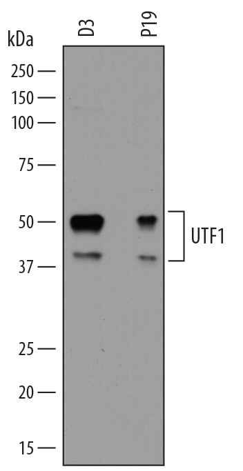
- Detection of Mouse UTF1 by Western Blot. Western blot shows lysates of D3 mouse embryonic stem cell line and P19 mouse embryonal carcinoma cell line. PVDF membrane was probed with 0.5 µg/mL of Sheep Anti-Mouse UTF1 Antigen Affinity-purified Polyclonal Antibody (Catalog # AF7819) followed by HRP-conjugated Anti-Sheep IgG Secondary Antibody (Catalog # HAF016). Specific bands were detected for UTF1 at approximately 50 and 40 kDa (as indicated). This experiment was conducted under reducing conditions and using Immunoblot Buffer Group 1.
Supportive validation
- Submitted by
- R&D Systems (provider)
- Main image
- Experimental details
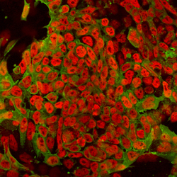
- UTF1 in D3 Mouse Stem Cells. UTF1 was detected in immersion fixed D3 mouse embryonic stem cell line using Sheep Anti-Mouse UTF1 Antigen Affinity-purified Polyclonal Antibody (Catalog # AF7819) at 10 µg/mL for 3 hours at room temperature. Cells were stained using the Northern-Lights™ 557-conjugated Anti-Sheep IgG Secondary Antibody (red; Catalog # NL010). Cells were double-stained using Mouse Anti-Human/Mouse SSEA-1 Monoclonal Antibody (Catalog # MAB2155) and the Northern-Lights™ 493-conjugated Anti-Mouse IgG Secondary Antibody (green; Catalog # NL009). Specific staining of UTF1 was localized to nuclei. View our protocol for Fluorescent ICC Staining of Cells on Coverslips.

Supportive validation
- Submitted by
- R&D Systems (provider)
- Main image

- Experimental details
- Detection of UTF1 in D3 Mouse Cell Line by Flow Cytometry. D3 mouse embryonic stem cell line untreated (panel A) or treated with 10 mM retinoic acid for 3 days (panel B) was stained with Sheep Anti-Mouse UTF1 Antigen Affinity-purified Polyclonal Antibody (Catalog # AF7819, filled histogram) or control antibody (Catalog # 5-001-A, open histogram), followed by Phycoerythrin-conjugated Anti-Sheep IgG Secondary Antibody (Catalog # F0126). UTF1 expression is decreased with retinoic acid treatment, as indicated in panel B. To facilitate intracellular staining, cells were fixed with paraformaldehyde and permeabilized with saponin.
