Explore
 Validate
 Learn
Learn Western blot
 Immunocytochemistry
Antibody data
- Antibody Data
- Antigen structure
- References [1]
- Comments [0]
- Validations
- Immunocytochemistry [1]
- Immunohistochemistry [1]
Submit
Validation data
Reference
Comment
Report error
- Product number
- PA3-752 - Provider product page

- Provider
- Invitrogen Antibodies
- Product name
- Anti-PRDX3 Polyclonal Antibody
- Antibody type
- Polyclonal
- Antigen
- Recombinant full-length protein
- Description
- PA3-753 detects Peroxiredoxin 4 protein in human samples. This antibody is specific for Prx 4 and shows no cross-reactivity with other Prx isoforms. PA3-753 has successfully been used in Western blot and immunocytochemistry procedures. By Western blot, this antibody detects an ~23 kDa and ~20 kDa protein representing pro-Prx 4 and mature Prx 4, respectively, in human PC3 cell lysate. The PA3-753 antigen is purified human recombinant Prx 4 from K562 cells.
- Reactivity
- Human
- Host
- Rabbit
- Isotype
- IgG
- Vial size
- 100 µL
- Concentration
- Lot Dependent
- Storage
- -20° C, Avoid Freeze/Thaw Cycles
Submitted references Nonredundant antioxidant defense by multiple two-cysteine peroxiredoxins in human prostate cancer cells.
Shen C, Nathan C
Molecular medicine (Cambridge, Mass.) 2002 Feb;8(2):95-102
Molecular medicine (Cambridge, Mass.) 2002 Feb;8(2):95-102
No comments: Submit comment
Supportive validation
- Submitted by
- Invitrogen Antibodies (provider)
- Main image
- Experimental details
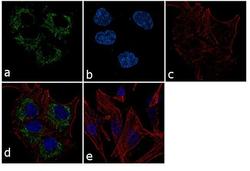
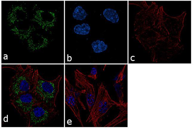
- Immunofluorescence analysis of Peroxiredoxin 3 was performed using 70% confluent log phase LNCaP cells. The cells were fixed with 4% paraformaldehyde for 10 minutes, permeabilized with 0.1% Triton™ X-100 for 10 minutes, and blocked with 1% BSA for 1 hour at room temperature. The cells were labeled with PRDX3 Rabbit Polyclonal Antibody (Product # PA3-752) at 1:250 dilution in 0.1% BSA and incubated for 3 hours at room temperature and then labeled with Goat anti-Rabbit IgG (H+L) Superclonal™ Secondary Antibody, Alexa Fluor® 488 conjugate (Product # A27034) at a dilution of 1:2000 for 45 minutes at room temperature (Panel a: green). Nuclei (Panel b: blue) were stained with SlowFade® Gold Antifade Mountant with DAPI (Product # S36938). F-actin (Panel c: red) was stained with Rhodamine Phalloidin (Product # R415, 1:300). Panel d represents the merged image showing cytoplasmic localization. Panel e shows the no primary antibody control. The images were captured at 60X magnification.
Supportive validation
- Submitted by
- Invitrogen Antibodies (provider)
- Main image
- Experimental details
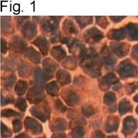
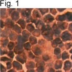
- Immunohistochemical staining of Prx 3 in TSU-Pr1 cells using Product # PA3-752.