 Explore
 Validate
 Learn
LearnNBP1-85123
antibody from Novus Biologicals
Targeting: HBS1L
DKFZp434g247, EF-1a, eRF3c, ERFS, HBS1, HSPC276, KIAA1038
 Western blot
 Immunocytochemistry
Antibody data
- Antibody Data
- Antigen structure
- References [1]
- Comments [0]
- Validations
- Western blot [2]
- Immunohistochemistry [1]
Submit
Validation data
Reference
Comment
Report error
- Product number
- NBP1-85123 - Provider product page

- Provider
- Novus Biologicals
- Proper citation
- Novus Cat#NBP1-85123, RRID:AB_11016830
- Product name
- Rabbit Polyclonal HBS1L Antibody
- Antibody type
- Polyclonal
- Description
- Immunogen affinity purified. Specificity of human, mouse, rat HBS1L antibody verified on a Protein Array containing target protein plus 383 other non-specific proteins.
- Reactivity
- Human, Mouse, Rat
- Host
- Rabbit
- Isotype
- IgG
- Vial size
- 0.1 ml
- Storage
- Store at 4C short term. Aliquot and store at -20C long term. Avoid freeze-thaw cycles.
Submitted references Systematic analysis of protein pools, isoforms, and modifications affecting turnover and subcellular localization.
Ahmad Y, Boisvert FM, Lundberg E, Uhlen M, Lamond AI
Molecular & cellular proteomics : MCP 2012 Mar;11(3):M111.013680
Molecular & cellular proteomics : MCP 2012 Mar;11(3):M111.013680
No comments: Submit comment
Supportive validation
- Submitted by
- Novus Biologicals (provider)
- Main image
- Experimental details
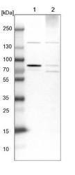
- Western Blot: HBS1L Antibody [NBP1-85123] - Lane 1: NIH-3T3 cell lysate (Mouse embryonic fibroblast cells). Lane 2: NBT-II cell lysate (Rat Wistar bladder tumor cells).

- Submitted by
- Novus Biologicals (provider)
- Main image

- Experimental details
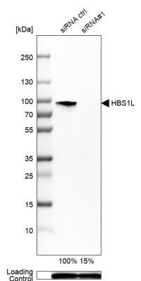
- Western Blot: HBS1L Antibody [NBP1-85123] - Western blot analysis in U-251MG cells transfected with control siRNA, target specific siRNA probe #1. Remaining relative intensity is presented. Loading control: Anti-GAPDH.
Supportive validation
- Submitted by
- Novus Biologicals (provider)
- Main image
- Experimental details
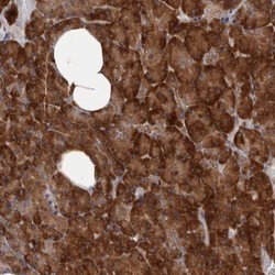
- Immunohistochemistry-Paraffin: HBS1L Antibody [NBP1-85123] - Staining of human pancreas shows strong cytoplasmic positivity in exocrine cells.
