 Explore
 Validate
 Learn
Learn Western blot
 Immunohistochemistry
Antibody data
- Antibody Data
- Antigen structure
- References [0]
- Comments [0]
- Validations
- Western blot [5]
- Immunocytochemistry [1]
Submit
Validation data
Reference
Comment
Report error
- Product number
- PA5-18478 - Provider product page

- Provider
- Invitrogen Antibodies
- Product name
- DDX6 Polyclonal Antibody
- Antibody type
- Polyclonal
- Antigen
- Synthetic peptide
- Description
- This antibody is predicted to react with canine, mouse and rat based on sequence homology. This antibody is tested in Peptide ELISA: antibody detection limit dilution 128,000.
- Reactivity
- Human
- Host
- Goat
- Isotype
- IgG
- Vial size
- 100 µg
- Concentration
- 0.5 mg/mL
- Storage
- -20° C, Avoid Freeze/Thaw Cycles
No comments: Submit comment
Supportive validation
- Submitted by
- Invitrogen Antibodies (provider)
- Main image

- Experimental details
- Western blot analysis of DDX6 by a DDX6 monoclonal antibody (Product # PA5-18478) at a concentration of 0.3 µg/mL. Daudi (A), Jurkat (B) and HepG2 (C) lysates (35µg protein in RIPA buffer). Primary incubation was 1 hour. Detected by chemiluminescence.

- Submitted by
- Invitrogen Antibodies (provider)
- Main image
- Experimental details
- Western Blot staining of Jurkat cell lysate using Product # PA5-18478 at a concentration of 0.3 µg/mL, the primary antibody incubation was 1 hour and the detection method was chemiluminescence.

- Submitted by
- Invitrogen Antibodies (provider)
- Main image

- Experimental details
- Western blot of HEK293 overexpressing Human DDX6 using Product # PA5-18478, mock transfection as a control in first lane.

- Submitted by
- Invitrogen Antibodies (provider)
- Main image

- Experimental details
- Knockdown of DDX6 was achieved by transfecting HeLa with DDX6 specific siRNAs (Silencer® select Product # s4010, s4011). Western blot analysis (Fig. a) was performed using whole cell extracts from the DDX6 knockdown cells (lane 3), non-specific scrambled siRNA transfected cells (lane 2) and untransfected cells (lane 1). The blot was probed with DDX6 Polyclonal Antibody (Product # PA5-18478, 1:1000 dilution) and Rabbit anti-Goat IgG (H+L) Superclonal™ Recombinant Secondary Antibody, HRP (Product # A27014, 1:4000 dilution). Densitometric analysis of this western blot is shown in histogram (Fig. b). Decrease in signal upon siRNA mediated knock down confirms that antibody is specific to DDX6.

- Submitted by
- Invitrogen Antibodies (provider)
- Main image
- Experimental details
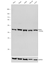
- Western blot was performed using DDX6 Polyclonal Antibody (Product # PA5-18478) and a 54 kDa band corresponding to DDX6 was observed across all the cell lines tested. Whole cell extracts (30 µg lysate) of HeLa (Lane 1), Daudi (Lane 2), Jurkat (Lane 3), Hep G2 (Lane 4) and A-431 (Lane 5) were electrophoresed using NuPAGE™ 10% Bis-Tris Protein Gel (Product # NP0302BOX). Resolved proteins were then transferred onto a nitrocellulose membrane (Product # IB23001) by iBlot® 2 Dry Blotting System (Product # IB21001). The blot was probed with the primary antibody (1:1000 dilution) and detected by chemiluminescence with Rabbit anti-Goat IgG (H+L), Superclonal™ Recombinant Secondary Antibody, HRP (Product # A27014, 1:4000 dilution) using the iBright FL 1000 (Product # A32752). Chemiluminescent detection was performed using Novex® ECL Chemiluminescent Substrate Reagent Kit (Product # WP20005).

Supportive validation
- Submitted by
- Invitrogen Antibodies (provider)
- Main image
- Experimental details
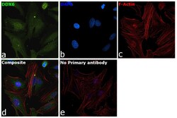
- Immunofluorescence analysis of DDX6 was performed using 70% confluent log phase HeLa cells. The cells were fixed with 4% Paraformaldehyde for 10 minutes, permeabilized with 0.1% Triton™ X-100 for 10 minutes, and blocked with 2% BSA for 1 hour at room temperature. The cells were labeled with DDX6 Polyclonal Antibody (Product # PA5-18478) at 1:100 dilution in 0.1% BSA, incubated at 4 degree Celsius overnight and then labeled with Donkey anti-Goat IgG (H+L) Highly Cross-Adsorbed Secondary Antibody, Alexa Fluor Plus 488 (Product # A32814) at 1:2000 dilution for 45 minutes at room temperature (Panel a: Green). Nuclei (Panel b: Blue) were stained with SlowFade® Gold Antifade Mountant with DAPI (Product # S36938). F-actin (Panel c: Red) was stained with Rhodamine Phalloidin (Product # R415, 1:300). Panel d represents the merged image showing cytoplasmic localization. Panel e represents control cells with no primary antibody to assess background. The images were captured at 60X magnification.
