Explore
 Validate
 Learn
Learn Western blot
 Blocking/Neutralizing
Antibody data
- Antibody Data
- Antigen structure
- References [3]
- Comments [0]
- Validations
- Western blot [1]
Submit
Validation data
Reference
Comment
Report error
- Product number
- AF2320 - Provider product page

- Provider
- R&D Systems
- Product name
- Human IL-36 gamma/IL-1F9 Antibody
- Antibody type
- Polyclonal
- Description
- Antigen Affinity-purified. Detects human IL-36 gamma/IL-1F9 in direct ELISAs and Western blots. In direct ELISAs, approximately 15% cross-reactivity with recombinant human (rh) IL-36 alpha is observed, approximately 5% cross-reactivity with rhIL-37 is observed, and less than 1% cross-reactivity with rhIL-1 alpha , rhIL-1 beta , rhIL-36Ra, and rhIL-36 beta is observed.
- Reactivity
- Human
- Host
- Goat
- Conjugate
- Unconjugated
- Antigen sequence
Q9NZH8- Isotype
- IgG
- Vial size
- 100 ug
- Concentration
- LYOPH
- Storage
- Use a manual defrost freezer and avoid repeated freeze-thaw cycles. 12 months from date of receipt, -20 to -70 °C as supplied. 1 month, 2 to 8 °C under sterile conditions after reconstitution. 6 months, -20 to -70 °C under sterile conditions after reconstitution.
Submitted references Application of IL-36 receptor antagonist weakens CCL20 expression and impairs recovery in the late phase of murine acetaminophen-induced liver injury.
The double-stranded RNA analogue polyinosinic-polycytidylic acid induces keratinocyte pyroptosis and release of IL-36γ.
IL-36γ/IL-1F9, an innate T-bet target in myeloid cells.
Scheiermann P, Bachmann M, Härdle L, Pleli T, Piiper A, Zwissler B, Pfeilschifter J, Mühl H
Scientific reports 2015 Feb 17;5:8521
Scientific reports 2015 Feb 17;5:8521
The double-stranded RNA analogue polyinosinic-polycytidylic acid induces keratinocyte pyroptosis and release of IL-36γ.
Lian LH, Milora KA, Manupipatpong KK, Jensen LE
The Journal of investigative dermatology 2012 May;132(5):1346-53
The Journal of investigative dermatology 2012 May;132(5):1346-53
IL-36γ/IL-1F9, an innate T-bet target in myeloid cells.
Bachmann M, Scheiermann P, Härdle L, Pfeilschifter J, Mühl H
The Journal of biological chemistry 2012 Dec 7;287(50):41684-96
The Journal of biological chemistry 2012 Dec 7;287(50):41684-96
No comments: Submit comment
Supportive validation
- Submitted by
- R&D Systems (provider)
- Main image

- Experimental details
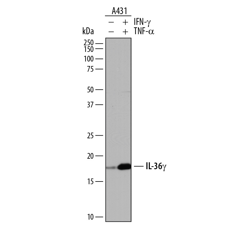
- Detection of Human IL-36 gamma/IL-1F9 by Western Blot. Western blot shows lysates of A431 human epithelial carcinoma cell line untreated (-) or treated (+) with 20 ng/mL Recombinant Human IFN-gamma (Catalog # 285-IF) and 20 ng/mL Recombinant Human TNF-alpha (Catalog # 210-TA) for 6 hours. PVDF membrane was probed with 0.5 µg/mL of Goat Anti-Human IL-36 gamma/IL-1F9 Antigen Affinity-purified Polyclonal Antibody (Catalog # AF2320) followed by HRP-conjugated Anti-Goat IgG Secondary Antibody (Catalog # HAF017). A specific band was detected for IL-36 gamma/IL-1F9 at approximately 18 kDa (as indicated). This experiment was conducted under reducing conditions and using Immunoblot Buffer Group 1.