Explore
 Validate
 Learn
LearnAF1078
antibody from R&D Systems
Targeting: IL36A
FIL1, FIL1E, IL-1F6, IL1(EPSILON), IL1F6, MGC129552, MGC129553
 Western blot
Antibody data
- Antibody Data
- Antigen structure
- References [2]
- Comments [0]
- Validations
- Western blot [1]
- Immunohistochemistry [1]
Submit
Validation data
Reference
Comment
Report error
- Product number
- AF1078 - Provider product page

- Provider
- R&D Systems
- Product name
- Human IL-36 alpha/IL-1F6 Antibody
- Antibody type
- Polyclonal
- Description
- Antigen Affinity-purified. Detects human IL-36 alpha/IL-1F6 in direct ELISAs and Western blots. In Western blots, less than 2% cross-reactivity with recombinant human (rh) rhIL-36 beta , rhIL-36 gamma , rhIL-1 alpha , rhIL-1 beta and rhIL-18 is observed.
- Reactivity
- Human
- Host
- Goat
- Conjugate
- Unconjugated
- Antigen sequence
Q9UHA7- Isotype
- IgG
- Vial size
- 100 ug
- Concentration
- LYOPH
- Storage
- Use a manual defrost freezer and avoid repeated freeze-thaw cycles. 12 months from date of receipt, -20 to -70 °C as supplied. 1 month, 2 to 8 °C under sterile conditions after reconstitution. 6 months, -20 to -70 °C under sterile conditions after reconstitution.
Submitted references Interleukin-36 receptor mediates the crosstalk between plasma cells and synovial fibroblasts.
IL-1F5, -F6, -F8, and -F9: a novel IL-1 family signaling system that is active in psoriasis and promotes keratinocyte antimicrobial peptide expression.
Schmitt V, Hahn M, Kästele V, Wagner O, Wiendl M, Derer A, Taddeo A, Hahne S, Radbruch A, Jäck HM, Schuh W, Mielenz D, Gay S, Schett G, Hueber AJ, Frey S
European journal of immunology 2017 Dec;47(12):2101-2112
European journal of immunology 2017 Dec;47(12):2101-2112
IL-1F5, -F6, -F8, and -F9: a novel IL-1 family signaling system that is active in psoriasis and promotes keratinocyte antimicrobial peptide expression.
Johnston A, Xing X, Guzman AM, Riblett M, Loyd CM, Ward NL, Wohn C, Prens EP, Wang F, Maier LE, Kang S, Voorhees JJ, Elder JT, Gudjonsson JE
Journal of immunology (Baltimore, Md. : 1950) 2011 Feb 15;186(4):2613-22
Journal of immunology (Baltimore, Md. : 1950) 2011 Feb 15;186(4):2613-22
No comments: Submit comment
Supportive validation
- Submitted by
- R&D Systems (provider)
- Main image
- Experimental details
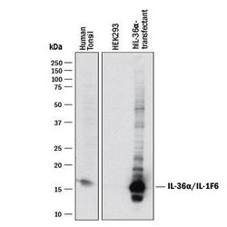
- Detection of Human IL-36 alpha/IL-1F6 by Western Blot. Western blot shows lysates of human tonsil tissue, HEK293 human embryonic kidney cell line either mock transfected or transfected with human IL-36 alpha/IL-1F6. PVDF membrane was probed with 1 µg/mL of Goat Anti-Human IL-36 alpha/IL-1F6 Antigen Affinity-purified Polyclonal Antibody (Catalog # AF1078) followed by HRP-conjugated Anti-Goat IgG Secondary Antibody (Catalog # HAF017). A specific band was detected for IL-36 alpha/IL-1F6 at approximately 16 kDa (as indicated). This experiment was conducted under reducing conditions and using Immunoblot Buffer Group 1.

Supportive validation
- Submitted by
- R&D Systems (provider)
- Main image

- Experimental details
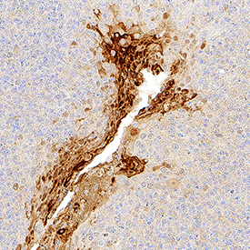
- IL-36 alpha/IL-1F6 in Human Tonsil. IL-36 alpha/IL-1F6 was detected in perfusion fixed paraffin-embedded sections of human tonsil using Goat Anti-Human IL-36 alpha/IL-1F6 Antigen Affinity-purified Polyclonal Antibody (Catalog # AF1078) at 1 µg/mL for 1 hour at room temperature followed by incubation with the Anti-Goat IgG VisUCyte™ HRP Polymer Antibody (Catalog # VC004). Tissue was stained using DAB (brown) and counterstained with hematoxylin (blue). Specific staining was localized to cytoplasm in lymphocytes. View our protocol for IHC Staining with VisUCyte HRP Polymer Detection Reagents.