Explore
 Validate
 Learn
Learn Western blot
Antibody data
- Antibody Data
- Antigen structure
- References [1]
- Comments [0]
- Validations
- Western blot [1]
- Immunohistochemistry [2]
- Other assay [1]
Submit
Validation data
Reference
Comment
Report error
- Product number
- PA5-119792 - Provider product page

- Provider
- Invitrogen Antibodies
- Product name
- INHBB Polyclonal Antibody
- Antibody type
- Polyclonal
- Antigen
- Synthetic peptide
- Description
- Positive Control: SK-Br-3 cell lysate, MCF-7 cell lysate, human uterus tissue, mouse brain tissue.
- Concentration
- 1 mg/mL
Submitted references INHBB is a novel prognostic biomarker and correlated with immune infiltrates in gastric cancer.
Yu W, He G, Zhang W, Ye Z, Zhong Z, Huang S
Frontiers in genetics 2022;13:933862
Frontiers in genetics 2022;13:933862
No comments: Submit comment
Supportive validation
- Submitted by
- Invitrogen Antibodies (provider)
- Main image

- Experimental details
- Western blot analysis of Inhibin beta B on different lysates. Proteins were transferred to a PVDF membrane and blocked with 5% BSA in PBS for 1 hour at room temperature. INHBB Polyclonal Antibody (Product # PA5-119792) at 1:500 was used in 5% BSA at room temperature for 2 hours. Goat Anti-Rabbit IgG - HRP Secondary Antibody at 1:5,000 dilution was used for 1 hour at room temperature. Positive control: Lane 1: SK-Br-3 cell lysate. Lane 2: MCF-7 cell lysate.

Supportive validation
- Submitted by
- Invitrogen Antibodies (provider)
- Main image
- Experimental details
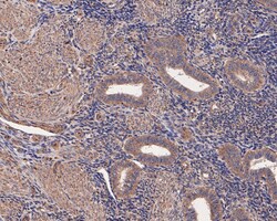
- Immunohistochemistry (Paraffin) analysis of paraffin-embedded human uterus tissue using INHBB Polyclonal Antibody (Product # PA5-119792). The section was pre-treated using heat mediated antigen retrieval with Tris-EDTA buffer (pH 8.0-8.4) for 20 minutes. The tissues were blocked in 5% BSA for 30 minutes at room temperature, washed with ddH2O and PBS, and then probed with the INHBB antibody at a dilution of 1:400 for 30 minutes at room temperature. The detection was performed using an HRP conjugated compact polymer system. DAB was used as the chromogen. Tissues were counterstained with hematoxylin and mounted with DPX.

- Submitted by
- Invitrogen Antibodies (provider)
- Main image
- Experimental details
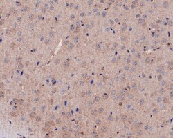
- Immunohistochemistry (Paraffin) analysis of paraffin-embedded mouse brain tissue using INHBB Polyclonal Antibody (Product # PA5-119792). The section was pre-treated using heat mediated antigen retrieval with Tris-EDTA buffer (pH 8.0-8.4) for 20 minutes. The tissues were blocked in 5% BSA for 30 minutes at room temperature, washed with ddH2O and PBS, and then probed with the INHBB antibody at a dilution of 1:400 for 30 minutes at room temperature. The detection was performed using an HRP conjugated compact polymer system. DAB was used as the chromogen. Tissues were counterstained with hematoxylin and mounted with DPX.

Supportive validation
- Submitted by
- Invitrogen Antibodies (provider)
- Main image
- Experimental details
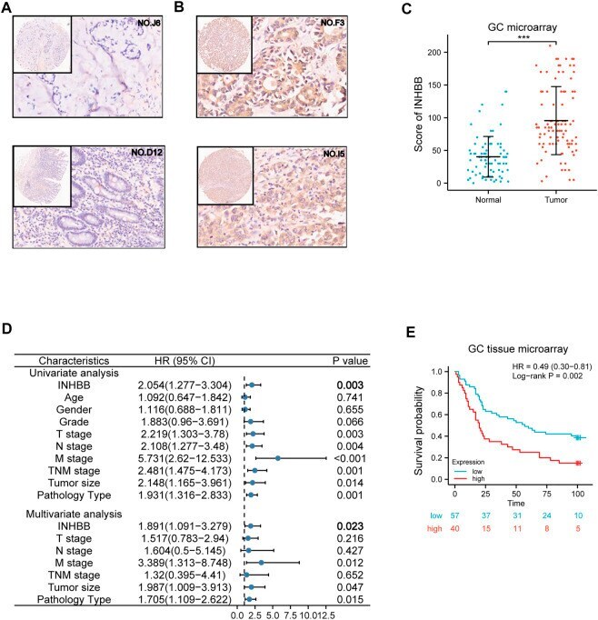
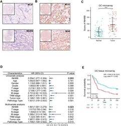
- FIGURE 5 Validation of the role of INHBB in GC. (A) IHC of normal tissues (4X and 20X scopes), (B) IHC of GC tissues (4X and 20X scopes), (C) differential expression of INHBB in GC tissues and normal tissues, (D) Cox regression analysis of INHBB and clinical features in GC, (E) Kaplan-Meier survival analyses of INHBB in GC patient data form GC tissue microarray.