 Explore
 Validate
 Learn
Learn Western blot
Antibody data
- Antibody Data
- Antigen structure
- References [1]
- Comments [0]
- Validations
- Western blot [4]
- Immunocytochemistry [2]
- Immunohistochemistry [3]
Submit
Validation data
Reference
Comment
Report error
- Product number
- GTX116314 - Provider product page

- Provider
- GeneTex
- Proper citation
- GeneTex Cat#GTX116314, RRID:AB_11176023
- Product name
- PHF10 antibody
- Antibody type
- Polyclonal
- Reactivity
- Human, Mouse, Rat
- Host
- Rabbit
Submitted references Vitamin D Switches BAF Complexes to Protect β Cells.
Wei Z, Yoshihara E, He N, Hah N, Fan W, Pinto AFM, Huddy T, Wang Y, Ross B, Estepa G, Dai Y, Ding N, Sherman MH, Fang S, Zhao X, Liddle C, Atkins AR, Yu RT, Downes M, Evans RM
Cell 2018 May 17;173(5):1135-1149.e15
Cell 2018 May 17;173(5):1135-1149.e15
No comments: Submit comment
Supportive validation
- Submitted by
- GeneTex (provider)
- Main image

- Experimental details
- Sample (30 ug of whole cell lysate) A: HeLa 10% SDS PAGE GTX116314 diluted at 1:1000

- Submitted by
- GeneTex (provider)
- Main image

- Experimental details
- PHF10 antibody detects PHF10 protein by western blot analysis.A.50 ?g mouse colon lysate/extract 10% SDS-PAGEPHF10 antibody (GTX116314) dilution: 1:1000 The HRP-conjugated anti-rabbit IgG antibody (GTX213110-01) was used to detect the primary antibody.

- Submitted by
- GeneTex (provider)
- Main image

- Experimental details
- HeLa whole cell and nuclear extracts (30 ?g) were separated by 10% SDS-PAGE, and the membrane was blotted with PHF10 antibody (GTX116314) diluted at 1:1000. The HRP-conjugated anti-rabbit IgG antibody (GTX213110-01) was used to detect the primary antibody.

- Submitted by
- GeneTex (provider)
- Main image

- Experimental details
- HeLa whole cell and nuclear extracts (30 ?g) were separated by 10% SDS-PAGE, and the membrane was blotted with PHF10 antibody (GTX116314) diluted at 1:1000.

Supportive validation
- Submitted by
- GeneTex (provider)
- Main image

- Experimental details
- Confocal immunofluorescence analysis (Olympus FV10i) of paraformaldehyde-fixed HeLa, using PHF10(GTX116314) antibody (Green) at 1:500 dilution. Alpha-tubulin filaments were labeled with GTX11304 (Red) at 1:2000.

- Submitted by
- GeneTex (provider)
- Main image

- Experimental details
- PHF10 antibody detects PHF10 protein at nucleus by immunofluorescent analysis.Sample: HeLa cells were fixed in 4% paraformaldehyde at RT for 15 min.Green: PHF10 protein stained by PHF10 antibody (GTX116314) diluted at 1:500.Red: Phalloidin, a cytoskeleton marker, diluted at 1:100.Blue: Hoechst 33342 staining.

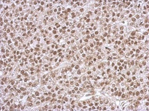
Supportive validation
- Submitted by
- GeneTex (provider)
- Main image

- Experimental details
- Immunohistochemical analysis of paraffin-embedded Hela xenograft, using PHF10(GTX116314) antibody at 1:500 dilution.
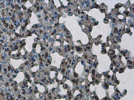
- Submitted by
- GeneTex (provider)
- Main image

- Experimental details
- PHF10 antibody detects PHF10 protein at nucleus in mouse lung by immunohistochemical analysis. Sample: Paraffin-embedded mouse lung. PHF10 antibody (GTX116314) diluted at 1:500.
- Submitted by
- GeneTex (provider)
- Main image

- Experimental details
- PHF10 antibody detects PHF10 protein at nucleus in rat lung by immunohistochemical analysis. Sample: Paraffin-embedded rat lung. PHF10 antibody (GTX116314) diluted at 1:500.
