 Explore
 Validate
 Learn
LearnNBP1-97593
antibody from Novus Biologicals
Targeting: NLRP1
CARD7, CLR17.1, DEFCAP, DKFZp586O1822, KIAA0926, NAC, NALP1, SLEV1, VAMAS1
 Western blot
 Immunocytochemistry
Antibody data
- Antibody Data
- Antigen structure
- References [2]
- Comments [0]
- Validations
- Western blot [1]
Submit
Validation data
Reference
Comment
Report error
- Product number
- NBP1-97593 - Provider product page

- Provider
- Novus Biologicals
- Proper citation
- Novus Cat#NBP1-97593, RRID:AB_11188534
- Product name
- Mouse Monoclonal NLRP1/NALP1 Antibody
- Antibody type
- Monoclonal
- Description
- Protein G purified. Recognizes the pyrin domain (PYD) of human NLRP1/NALP1..
- Reactivity
- Human
- Host
- Mouse
- Isotype
- IgG
- Vial size
- 0.05 mg
- Concentration
- 1.0 mg/ml
- Storage
- Aliquot and store at -20C or -80C. Avoid freeze-thaw cycles.
Submitted references Expression of Th17- and Treg-specific transcription factors in vitiligo patients.
Inflammasome components NALP 1 and 3 show distinct but separate expression profiles in human tissues suggesting a site-specific role in the inflammatory response.
Bhardwaj S, Rani S, Kumaran MS, Bhatia A, Parsad D
International journal of dermatology 2020 Apr;59(4):474-481
International journal of dermatology 2020 Apr;59(4):474-481
Inflammasome components NALP 1 and 3 show distinct but separate expression profiles in human tissues suggesting a site-specific role in the inflammatory response.
Kummer JA, Broekhuizen R, Everett H, Agostini L, Kuijk L, Martinon F, van Bruggen R, Tschopp J
The journal of histochemistry and cytochemistry : official journal of the Histochemistry Society 2007 May;55(5):443-52
The journal of histochemistry and cytochemistry : official journal of the Histochemistry Society 2007 May;55(5):443-52
No comments: Submit comment
Supportive validation
- Submitted by
- Novus Biologicals (provider)
- Main image
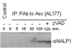
- Experimental details
- Western Blot: NLRP1/NALP1 Antibody (Nalpy1-4) [NBP1-97593] - Analysis of the time course of assembly of the inflammasome using MAb to NLRP1/NALP1 (Nalpy1-4) in THP-1 macrophages. Method: Assembly of the inflammasome was induced by shifting the temperature to 30C after hypotonic lysis. THP-1 cell extracts were immunoprecipitated using PAb to Asc (AL177) and run on SDS-PAGE. NLRP1/NALP1 was detected by MAb to NALP1 (Nalpy1-4) at 1:1000 dilution. Anti-mouse IgG coupled horse radish peroxidase was used at 1:5000 dilution for ECL detection. For more information about the 'time course assembly of inflammasome' method see F. Martinon; Mol. Cell. 10, 417 (2002).
