Explore
 Validate
 Learn
Learn Western blot
Antibody data
- Antibody Data
- Antigen structure
- References [3]
- Comments [0]
- Validations
- Western blot [1]
Submit
Validation data
Reference
Comment
Report error
- Product number
- PAB10006 - Provider product page

- Provider
- Abnova Corporation
- Proper citation
- Abnova Corporation Cat#PAB10006, RRID:AB_1707660
- Product name
- ECT2 (phospho T790) polyclonal antibody
- Antibody type
- Polyclonal
- Description
- Rabbit polyclonal antibody raised against synthetic phosphopeptide of ECT2.
- Storage
- Store at 4°C. For long term storage store at -20°C.Aliquot to avoid repeated freezing and thawing.
Submitted references MgcRacGAP controls the assembly of the contractile ring and the initiation of cytokinesis.
Inhibition of cyclin-dependent kinase 1 induces cytokinesis without chromosome segregation in an ECT2 and MgcRacGAP-dependent manner.
The tandem BRCT domains of Ect2 are required for both negative and positive regulation of Ect2 in cytokinesis.
Zhao WM, Fang G
Proceedings of the National Academy of Sciences of the United States of America 2005 Sep 13;102(37):13158-63
Proceedings of the National Academy of Sciences of the United States of America 2005 Sep 13;102(37):13158-63
Inhibition of cyclin-dependent kinase 1 induces cytokinesis without chromosome segregation in an ECT2 and MgcRacGAP-dependent manner.
Niiya F, Xie X, Lee KS, Inoue H, Miki T
The Journal of biological chemistry 2005 Oct 28;280(43):36502-9
The Journal of biological chemistry 2005 Oct 28;280(43):36502-9
The tandem BRCT domains of Ect2 are required for both negative and positive regulation of Ect2 in cytokinesis.
Kim JE, Billadeau DD, Chen J
The Journal of biological chemistry 2005 Feb 18;280(7):5733-9
The Journal of biological chemistry 2005 Feb 18;280(7):5733-9
No comments: Submit comment
Supportive validation
- Submitted by
- Abnova Corporation (provider)
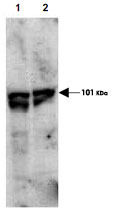
- Main image
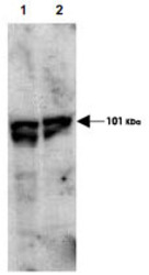
- Experimental details
- Western blot using ECT2 (phospho T790) polyclonal antibody (Cat # PAB10006) showsdetection of endogenous phospho-ECT2 (arrowhead) present in cell lysates from interphase (Lane 1) and mitotic (Lane 2) HeLa cells.Despite specific staining of interphase cells, this reagent is believed to be phospho specific based on ELISA resultsusing both phosphorylated and non-phosphorylated immunizing peptide.After SDS-PAGE and transfer, the membrane was probed with the primary antibody diluted to 1 : 1,000.Personal Communication, Toru Miki,CCR-NCI, Bethesda, MD.