 Explore
 Validate
 Learn
Learn Western blot
Antibody data
- Antibody Data
- Antigen structure
- References [1]
- Comments [0]
- Validations
- Western blot [1]
- Immunocytochemistry [3]
- Immunohistochemistry [1]
Submit
Validation data
Reference
Comment
Report error
- Product number
- GTX115575 - Provider product page

- Provider
- GeneTex
- Proper citation
- GeneTex Cat#GTX115575, RRID:AB_10619841
- Product name
- Cpn10 antibody
- Antibody type
- Polyclonal
- Reactivity
- Human, Mouse
- Host
- Rabbit
Submitted references Comparative analysis of Hsp10 and Hsp90 expression in healthy mucosa and adenocarcinoma of the large bowel.
Rappa F, Sciume C, Lo Bello M, Bavisotto CC, Marino Gammazza A, Barone R, Campanella C, David S, Carini F, Zarcone F, Rizzuto S, Lena A, Tomasello G, Uzzo ML, Spatola GF, Bonaventura G, Leone A, Gerbino A, Cappello F, Bucchieri F, Zummo G, Farina F
Anticancer research 2014 Aug;34(8):4153-9
Anticancer research 2014 Aug;34(8):4153-9
No comments: Submit comment
Supportive validation
- Submitted by
- GeneTex (provider)
- Main image
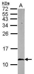
- Experimental details
- Sample (50 ug of whole cell lysate) A: mouse brain 15% SDS PAGE GTX115575 diluted at 1:1000

Supportive validation
- Submitted by
- GeneTex (provider)
- Main image
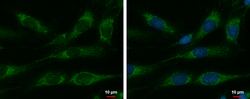
- Experimental details
- Cpn10 antibody detects Cpn10 protein at mitochondria by immunofluorescent analysis.Sample: SKNSH cells were fixed in 2% paraformaldehyde/culture medium at 37¢J for 15 min.Green: Cpn10 protein stained by Cpn10 antibody (GTX115575) diluted at 1:500.Blue: Hoechst 33342 staining.

- Submitted by
- GeneTex (provider)
- Main image

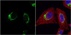
- Experimental details
- Cpn10 antibody detects Cpn10 protein at mitochondria by immunofluorescent analysis.Sample: HeLa cells were fixed in 4% paraformaldehyde at RT for 15 min.Green: Cpn10 protein stained by Cpn10 antibody (GTX115575) diluted at 1:100.Red: alpha Tubulin, a cytoskeleton marker, stained by alpha Tubulin antibody [B-5-1-2] (GTX11304) diluted at 1:10000.Blue: Hoechst 33342 staining.

- Submitted by
- GeneTex (provider)
- Main image
- Experimental details
- Cpn10 antibody detects Cpn10 protein at cytoplasm by immunofluorescent analysis.Sample: U2OS cells were fixed in 4% paraformaldehyde at RT for 15 min.Green: Cpn10 protein stained by Cpn10 antibody (GTX115575) diluted at 1:200.Red: alpha Tubulin, a cytoskeleton marker, stained by alpha Tubulin antibody [GT114] (GTX628802) diluted at 1:1000.Blue: Hoechst 33342 staining.

Supportive validation
- Submitted by
- GeneTex (provider)
- Main image
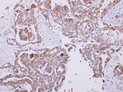
- Experimental details
- Cpn10 antibody detects Cpn10 protein at cytoplasm on human lung carcinoma by immunohistochemical analysis. Sample: Paraffin-embedded lung carcinoma. Cpn10 antibody (GTX115575) dilution: 1:250.
