Explore
 Validate
 Learn
Learn Western blot
 Flow cytometry
Antibody data
- Antibody Data
- Antigen structure
- References [2]
- Comments [0]
- Validations
- Flow cytometry [1]
Submit
Validation data
Reference
Comment
Report error
- Product number
- BAF2154 - Provider product page

- Provider
- Novus Biologicals
- Product name
- Goat Polyclonal B7-H4 Antibody
- Antibody type
- Polyclonal
- Description
- Antigen Affinity-purified. Detects mouse B7-H4 in Western blots. In Western blots, less than 1% cross-reactivity with recombinant mouse (rm) B7-1, rmB7-2, rmB7-H1, rmB7-H2, rmB7-H3, and rmPD-L2 is observed.
- Reactivity
- Mouse
- Host
- Goat
- Conjugate
- Biotin
- Isotype
- IgG
- Vial size
- 50 ug
- Concentration
- LYOPH
- Storage
- Use a manual defrost freezer and avoid repeated freeze-thaw cycles. 12 months from date of receipt, -20 to -70 degreesC as supplied. 1 month, 2 to 8 degreesC under sterile conditions after reconstitution. 6 months, -20 to -70 degreesC under sterile conditions after reconstitution.
Submitted references Tissue-expressed B7x affects the immune response to and outcome of lethal pulmonary infection.
B7x in the periphery abrogates pancreas-specific damage mediated by self-reactive CD8 T cells.
Hofmeyer KA, Scandiuzzi L, Ghosh K, Pirofski LA, Zang X
Journal of immunology (Baltimore, Md. : 1950) 2012 Sep 15;189(6):3054-63
Journal of immunology (Baltimore, Md. : 1950) 2012 Sep 15;189(6):3054-63
B7x in the periphery abrogates pancreas-specific damage mediated by self-reactive CD8 T cells.
Lee JS, Scandiuzzi L, Ray A, Wei J, Hofmeyer KA, Abadi YM, Loke P, Lin J, Yuan J, Serreze DV, Allison JP, Zang X
Journal of immunology (Baltimore, Md. : 1950) 2012 Oct 15;189(8):4165-74
Journal of immunology (Baltimore, Md. : 1950) 2012 Oct 15;189(8):4165-74
No comments: Submit comment
Supportive validation
- Submitted by
- Novus Biologicals (provider)
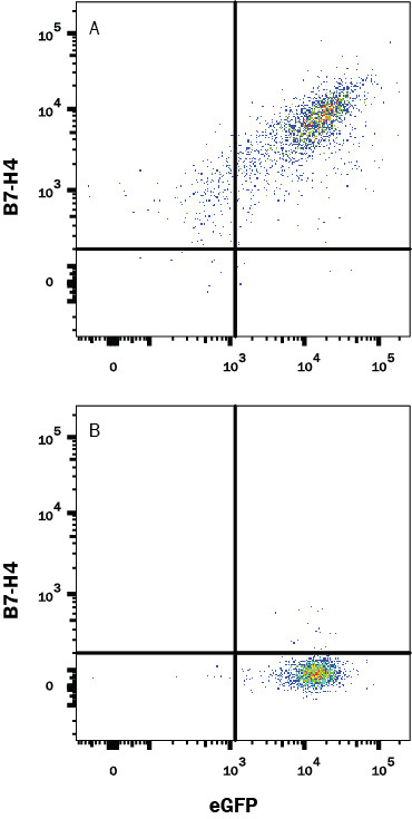
- Main image

- Experimental details
- Detection of B7-H4 in HEK293 Human Cell Line Transfected with Mouse B7-H4 and eGFP by Flow Cytometry. Detection of B7-H4 in HEK293 Human Cell Line Transfected with Mouse B7-H4 and eGFP by Flow Cytometry.