 Explore
 Validate
 Learn
Learn Western blot
Antibody data
- Antibody Data
- Antigen structure
- References [7]
- Comments [0]
- Validations
- Western blot [7]
- Immunohistochemistry [1]
- Other assay [2]
Submit
Validation data
Reference
Comment
Report error
- Product number
- PA5-27828 - Provider product page

- Provider
- Invitrogen Antibodies
- Product name
- Collagen III Polyclonal Antibody
- Antibody type
- Polyclonal
- Antigen
- Recombinant protein fragment
- Description
- Recommended positive controls: HeLa, COL3A1 shRNA-transfected HeLa, SK-N-SH, SK-N-AS, Neuro2A, NIH-3T3, BCL-1, Raw264.7, C2C12, Mouse ESC, PC-12, Rat2, rat liver.
- Concentration
- 0.46 mg/mL
Submitted references Downregulation of senescence-associated secretory phenotype by knockdown of secreted frizzled-related protein 4 contributes to the prevention of skin aging.
Effects and Mechanism of Particulate Matter on Tendon Healing Based on Integrated Analysis of DNA Methylation and RNA Sequencing Data in a Rat Model.
Impaired non-canonical transforming growth factor-β signalling prevents profibrotic phenotypes in cultured peptidylarginine deiminase 4-deficient murine cardiac fibroblasts.
Chronic High Glucose Concentration Induces Inflammatory and Remodeling Changes in Valvular Endothelial Cells and Valvular Interstitial Cells in a Gelatin Methacrylate 3D Model of the Human Aortic Valve.
Cathepsin B deficiency ameliorates liver lipid deposition, inflammatory cell infiltration, and fibrosis after diet-induced nonalcoholic steatohepatitis.
ACVR1(R206H) FOP mutation alters mechanosensing and tissue stiffness during heterotopic ossification.
Morphological and Molecular Changes in Juvenile Normal Human Fibroblasts Exposed to Simulated Microgravity.
Takaya K, Asou T, Kishi K
Aging 2022 Sep 7;14(20):8167-8178
Aging 2022 Sep 7;14(20):8167-8178
Effects and Mechanism of Particulate Matter on Tendon Healing Based on Integrated Analysis of DNA Methylation and RNA Sequencing Data in a Rat Model.
Lee SY, Lee MH, Jo SK, Yoo IH, Sarankhuu BE, Kim HJ, Kang YE, Lee SE, Kim TY, Park MH, Lee CS, Han SY, Moon JH, Jung JY, Hong GL, Yoo NJ, Yoon ES, Choi JK, Won HR, Son JW, Song JH
International journal of molecular sciences 2022 Jul 25;23(15)
International journal of molecular sciences 2022 Jul 25;23(15)
Impaired non-canonical transforming growth factor-β signalling prevents profibrotic phenotypes in cultured peptidylarginine deiminase 4-deficient murine cardiac fibroblasts.
Akboua H, Eghbalzadeh K, Keser U, Wahlers T, Paunel-Görgülü A
Journal of cellular and molecular medicine 2021 Oct;25(20):9674-9684
Journal of cellular and molecular medicine 2021 Oct;25(20):9674-9684
Chronic High Glucose Concentration Induces Inflammatory and Remodeling Changes in Valvular Endothelial Cells and Valvular Interstitial Cells in a Gelatin Methacrylate 3D Model of the Human Aortic Valve.
Ciortan L, Macarie RD, Cecoltan S, Vadana M, Tucureanu MM, Mihaila AC, Droc I, Butoi E, Manduteanu I
Polymers 2020 Nov 25;12(12)
Polymers 2020 Nov 25;12(12)
Cathepsin B deficiency ameliorates liver lipid deposition, inflammatory cell infiltration, and fibrosis after diet-induced nonalcoholic steatohepatitis.
Fang W, Deng Z, Benadjaoud F, Yang C, Shi GP
Translational research : the journal of laboratory and clinical medicine 2020 Aug;222:28-40
Translational research : the journal of laboratory and clinical medicine 2020 Aug;222:28-40
ACVR1(R206H) FOP mutation alters mechanosensing and tissue stiffness during heterotopic ossification.
Haupt J, Stanley A, McLeod CM, Cosgrove BD, Culbert AL, Wang L, Mourkioti F, Mauck RL, Shore EM
Molecular biology of the cell 2019 Jan 1;30(1):17-29
Molecular biology of the cell 2019 Jan 1;30(1):17-29
Morphological and Molecular Changes in Juvenile Normal Human Fibroblasts Exposed to Simulated Microgravity.
Buken C, Sahana J, Corydon TJ, Melnik D, Bauer J, Wehland M, Krüger M, Balk S, Abuagela N, Infanger M, Grimm D
Scientific reports 2019 Aug 15;9(1):11882
Scientific reports 2019 Aug 15;9(1):11882
No comments: Submit comment
Supportive validation
- Submitted by
- Invitrogen Antibodies (provider)
- Main image
- Experimental details
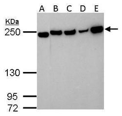
- Western blot analysis of COL3A1 using A) 30 µg Neuro2A whole lysate (B) 30 µg NIH-3T3 whole cell lysate (C) 30 µg BCL-1 whole cell lysate (D) 30 µg Raw264.7 whole cell lysate and E) 30 µg C2C12 whole cell lysate. Samples were loaded onto a 5% SDS-PAGE gel and probed with a COL3A1 polyclonal antibody (Product # PA5-27828) at a dilution of 1:1000.

- Submitted by
- Invitrogen Antibodies (provider)
- Main image

- Experimental details
- Western blot analysis of COL3A1 using A) 30 µg PC-12 whole cell extract and B) 30 µg Rat2 whole cell extract. Samples were loaded onto a 5% SDS-PAGE gel and probed with a COL3A1 polyclonal antibody (Product # PA5-27828) at a dilution of 1:1000.

- Submitted by
- Invitrogen Antibodies (provider)
- Main image

- Experimental details
- Western blot analysis of COL3A1 using 50 µg rat liver lysate. Samples were loaded onto a 5% SDS-PAGE gel and probed with a COL3A1 polyclonal antibody (Product # PA5-27828) at a dilution of 1:1000.

- Submitted by
- Invitrogen Antibodies (provider)
- Main image

- Experimental details
- Western blot analysis of COL3A1 using 30 µg of HeLa lysate. Samples were loaded onto a 5% SDS-PAGE gel and probed with a COL3A1 polyclonal antibody (Product # PA5-27828) at a dilution of 1:1000.

- Submitted by
- Invitrogen Antibodies (provider)
- Main image

- Experimental details
- Western Blot analysis of Collagen III was performed by separating 30 µg of mouse tissue extracts by 5% SDS-PAGE. Proteins were transferred to a membrane and probed with a Collagen III Polyclonal Antibody (Product # PA5-27828) at a dilution of 1:1000. The HRP-conjugated anti-rabbit IgG antibody was used to detect the primary antibody.

- Submitted by
- Invitrogen Antibodies (provider)
- Main image

- Experimental details
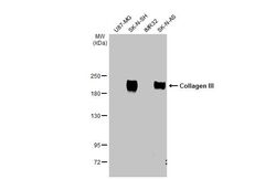
- Western Blot analysis of Collagen III was performed by separating 30 µg of various whole cell extracts by 5% SDS-PAGE. Proteins were transferred to a membrane and probed with a Collagen III Polyclonal Antibody (Product # PA5-27828) at a dilution of 1:5000 and a HRP-conjugated anti-rabbit IgG secondary antibody.

- Submitted by
- Invitrogen Antibodies (provider)
- Main image
- Experimental details
- Western Blot using Collagen III Polyclonal Antibody (Product # PA5-27828). Various extracts (30 µg) were separated by 5% SDS-PAGE, and the membrane was blotted with Collagen III Polyclonal Antibody (Product # PA5-27828) diluted at 1:1,000. The HRP-conjugated anti-rabbit IgG antibody was used to detect the primary antibody.

Supportive validation
- Submitted by
- Invitrogen Antibodies (provider)
- Main image
- Experimental details
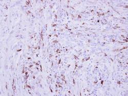
- Immunohistochemical analysis of paraffin-embedded human PAPILLARY CA_STROMAL CELLS, using Collagen III (Product # PA5-27828) antibody at 1:250 dilution. Antigen Retrieval: EDTA based buffer, pH 8.0, 15 min.

Supportive validation
- Submitted by
- Invitrogen Antibodies (provider)
- Main image
- Experimental details
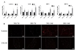
- Figure 6 ( A ) Gene expression of extracellular matrix (ECM) proteins and MMPs (MMP-1 and MMP-13) in VEC and VIC cultured in 3D in HG versus NG glucose exposure, as evaluated by real-time PCR. The mRNA of investigated molecules was normalized to actin mRNA. The results are presented as fold change versus NG condition for every type of cell and investigated time. N = 3, * p < 0.5, ** p < 0.01. ( B ) Identification of ECM protein in 3D constructs with VEC and VIC, grown in NG or HG for 7 and 14 days. Representative images of laminin alpha4 chain and collagen III in sections obtained from 3D constructs grown in NG and HG for 7 and 14 days. The images were taken with an 10x objective, with filter for DAPI (nuclei-cyan) and filter Alexa 594 (laminin and collagen III-red).

- Submitted by
- Invitrogen Antibodies (provider)
- Main image

- Experimental details
- FIGURE 2 Expression of fibrosis-related markers in PAD4 -/- hearts. (A) The expression of fibrosis-related genes in WT ( n = 7) and PAD4 -/- ( n = 10) hearts was analysed by real-time PCR. (B) alpha-SMA expression was detected by immunofluorescence and was found to be restricted to vascular cells ( white arrow heads ). One representative image of three stained hearts is depicted. Scale bar indicates 100 um. n = 5/group. (C) Collagen I and III protein expression in cardiac tissue of WT and PAD4 -/- mice. n = 7. (D) Representative images of heart sections from WT and PAD4-deficient mice stained with Masson trichrome under baseline conditions. * p < 0.05
