 Explore
 Validate
 Learn
Learn Western blot
Antibody data
- Antibody Data
- Antigen structure
- References [3]
- Comments [0]
- Validations
- Western blot [3]
- Immunocytochemistry [2]
- Immunohistochemistry [1]
Submit
Validation data
Reference
Comment
Report error
- Product number
- GTX113542 - Provider product page

- Provider
- GeneTex
- Proper citation
- GeneTex Cat#GTX113542, RRID:AB_2037923
- Product name
- RPS6 antibody
- Antibody type
- Polyclonal
- Reactivity
- Human, Mouse, Zebrafish
- Host
- Rabbit
Submitted references Rhapontigenin inhibits TGF-β-mediated epithelial‑mesenchymal transition via the PI3K/AKT/mTOR pathway and is not associated with HIF-1α degradation.
Cdc6 cooperates with c-Myc to promote genome instability and epithelial to mesenchymal transition EMT in zebrafish.
Activation of the mTOR pathway by low levels of xenoestrogens in breast epithelial cells from high-risk women.
Yeh YH, Wang SW, Yeh YC, Hsiao HF, Li TK
Oncology reports 2016 May;35(5):2887-95
Oncology reports 2016 May;35(5):2887-95
Cdc6 cooperates with c-Myc to promote genome instability and epithelial to mesenchymal transition EMT in zebrafish.
Chen CH, Lin DS, Cheng CW, Lin CJ, Lo YK, Yen CC, Lee AY, Hsiao CD
Oncotarget 2014 Aug 15;5(15):6300-11
Oncotarget 2014 Aug 15;5(15):6300-11
Activation of the mTOR pathway by low levels of xenoestrogens in breast epithelial cells from high-risk women.
Goodson WH 3rd, Luciani MG, Sayeed SA, Jaffee IM, Moore DH 2nd, Dairkee SH
Carcinogenesis 2011 Nov;32(11):1724-33
Carcinogenesis 2011 Nov;32(11):1724-33
No comments: Submit comment
Supportive validation
- Submitted by
- GeneTex (provider)
- Main image

- Experimental details
- Sample (30 ug of whole cell lysate) A: 293T B: A431 (GTX27909) 12% SDS PAGE GTX113542 diluted at 1:1000
- Validation comment
- WB

- Submitted by
- GeneTex (provider)
- Main image

- Experimental details
- Sample (50 ?g of whole cell lysate) A: mouse brain 12% SDS PAGE GTX113542 diluted at 1:500 The HRP-conjugated anti-rabbit IgG antibody (GTX213110-01) was used to detect the primary antibody.

- Submitted by
- GeneTex (provider)
- Main image

- Experimental details
- Various whole cell extracts (30 ?g) were separated by 12% SDS-PAGE, and the membrane was blotted with RPS6 antibody (GTX113542) diluted at 1:500. The HRP-conjugated anti-rabbit IgG antibody (GTX213110-01) was used to detect the primary antibody.

Supportive validation
- Submitted by
- GeneTex (provider)
- Main image

- Experimental details
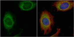
- Confocal immunofluorescence analysis (Olympus FV10i) of paraformaldehyde-fixed A431, using RPS6(GTX113542) antibody (Green) at 1:500 dilution. Alpha-tubulin filaments were labeled with GTX11304 (Red) at 1:500.

- Submitted by
- GeneTex (provider)
- Main image
- Experimental details
- RPS6 antibody detects RPS6 protein at cytoplasm by immunofluorescent analysis.Sample: HeLa cells were fixed in 4% paraformaldehyde at RT for 15 min.Green: RPS6 protein stained by RPS6 antibody (GTX113542) diluted at 1:500.Red: alpha Tubulin, a cytoskeleton marker, stained by alpha Tubulin antibody [GT114] (GTX628802) diluted at 1:500.Blue: Hoechst 33342 staining.

Supportive validation
- Submitted by
- GeneTex (provider)
- Main image
- Experimental details
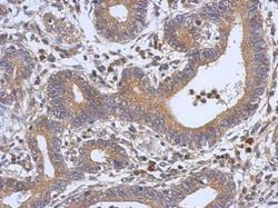
- Immunohistochemical analysis of paraffin-embedded human colon carcinoma, using RPS6(GTX113542) antibody at 1:500 dilution.
