 Explore
 Validate
 Learn
Learn Western blot
 Immunocytochemistry
Antibody data
- Antibody Data
- Antigen structure
- References [1]
- Comments [0]
- Validations
- Western blot [8]
- Immunocytochemistry [2]
- Immunoprecipitation [1]
Submit
Validation data
Reference
Comment
Report error
- Product number
- GTX629818 - Provider product page

- Provider
- GeneTex
- Product name
- IDH1 antibody [GT1521]
- Antibody type
- Monoclonal
- Reactivity
- Human, Mouse, Rat
- Host
- Mouse
Submitted references A clinical drug library screen identifies clobetasol propionate as an NRF2 inhibitor with potential therapeutic efficacy in KEAP1 mutant lung cancer.
Choi EJ, Jung BJ, Lee SH, Yoo HS, Shin EA, Ko HJ, Chang S, Kim SY, Jeon SM
Oncogene 2017 Sep 14;36(37):5285-5295
Oncogene 2017 Sep 14;36(37):5285-5295
No comments: Submit comment
Enhanced validation
Supportive validation
- Submitted by
- GeneTex (provider)
- Enhanced method
- Genetic validation
- Main image

- Experimental details
- Non-transfected (¡V) and transfected (+) HepG2 whole cell extracts (30 ?g) were separated by 10% SDS-PAGE, and the membrane was blotted with IDH1 antibody [GT1521] (GTX629818) diluted at 1:4000. The HRP-conjugated anti-mouse IgG antibody (GTX213111-01) was used to detect the primary antibody.

Supportive validation
- Submitted by
- GeneTex (provider)
- Main image

- Experimental details
- IDH1 antibody [GT1521] detects IDH1 protein by western blot analysis.A. 30 ?g 293T whole cell lysate/extractB. 30 ?g A431 whole cell lysate/extractC. 30 ?g HeLa whole cell lysate/extractD. 30 ?g HepG2 whole cell lysate/extract10% SDS-PAGEIDH1 antibody [GT1521] (GTX629818) dilution: 1:1000The HRP-conjugated anti-mouse IgG antibody (GTX213111-01) was used to detect the primary antibody.

- Submitted by
- GeneTex (provider)
- Main image

- Experimental details
- IDH1 antibody [GT1521] detects IDH1 protein by western blot analysis.A. 30 ?g PC-12 whole cell lysate/extractB. 30 ?g Rat-2 whole cell lysate/extract10% SDS-PAGEIDH1 antibody [GT1521] (GTX629818) dilution: 1:1000The HRP-conjugated anti-mouse IgG antibody (GTX213111-01) was used to detect the primary antibody.

- Submitted by
- GeneTex (provider)
- Main image

- Experimental details
- IDH1 antibody [GT1521] detects IDH1 protein by western blot analysis.A. 30 ?g Neuro2A whole cell lysate/extractB. 30 ?g GL261 whole cell lysate/extractC. 30 ?g C8D30 whole cell lysate/extractD. 30 ?g NIH-3T3 whole cell lysate/extract E. 30 ?g Raw264.7 whole cell lysate/extractF. 30 ?g C2C12 whole cell lysate/extract10% SDS-PAGEIDH1 antibody [GT1521] (GTX629818) dilution: 1:1000The HRP-conjugated anti-mouse IgG antibody (GTX213111-01) was used to detect the primary antibody.

- Submitted by
- GeneTex (provider)
- Main image

- Experimental details
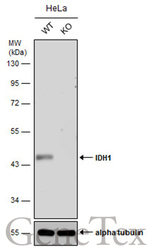
- Wild-type (WT) and IDH1 knockout (KO) HeLa cell extracts (30 ?g) were separated by 10% SDS-PAGE, and the membrane was blotted with IDH1 antibody [GT1521] (GTX629818) diluted at 1:500. The HRP-conjugated anti-mouse IgG antibody (GTX213111-01) was used to detect the primary antibody.

- Submitted by
- GeneTex (provider)
- Main image

- Experimental details
- Non-transfected (¡V) and transfected (+) HepG2 whole cell extracts (30 ?g) were separated by 10% SDS-PAGE, and the membrane was blotted with IDH1 antibody [GT1521] (GTX629818) diluted at 1:4000. The HRP-conjugated anti-mouse IgG antibody (GTX213111-01) was used to detect the primary antibody.

- Submitted by
- GeneTex (provider)
- Main image
- Experimental details
- Wild-type (WT) and IDH1 knockout (KO) HeLa cell extracts (30 ?g) were separated by 10% SDS-PAGE, and the membrane was blotted with IDH1 antibody [GT1521] (GTX629818) diluted at 1:500. The HRP-conjugated anti-mouse IgG antibody (GTX213111-01) was used to detect the primary antibody.

- Submitted by
- GeneTex (provider)
- Main image

- Experimental details
- Various whole cell extracts (30 ?g) were separated by 7.5% SDS-PAGE, and the membranes were blotted with IDH1 antibody [GT1521] (GTX629818) diluted at 1:1000 and competitor's antibody (CST#8137) diluted at 1:1000. The HRP-conjugated anti-mouse IgG antibody (GTX213111-01) was used to detect the primary antibody.

Supportive validation
- Submitted by
- GeneTex (provider)
- Main image

- Experimental details
- IDH1 antibody [GT1521] detects IDH1 protein at cytoplasm by immunofluorescent analysis.Sample: A431 cells were fixed in ice-cold MeOH for 5 min.Green: IDH1 protein stained by IDH1 antibody [GT1521] (GTX629818) diluted at 1:200.Blue: Hoechst 33342 staining.

- Submitted by
- GeneTex (provider)
- Main image

- Experimental details
- IDH1 antibody [GT1521] detects IDH1 protein at cytoplasm by immunofluorescent analysis.Sample: HepG2 cells were fixed in 4% paraformaldehyde at RT for 15 min.Green: IDH1 protein stained by IDH1 antibody [GT1521] (GTX629818) diluted at 1:500.Blue: Hoechst 33342 staining.

Supportive validation
- Submitted by
- GeneTex (provider)
- Main image

- Experimental details
- IDH-1 antibody immunoprecipitates IDH-1 protein in IP experiments. IP Sample: HepG2 whole cell lysate/extract A : 30 £gg whole cell lysate/extract of IDH1 protein expressing HepG2 cells B : Control with 2.5 £gg of pre-immune mouse IgG C : Immunoprecipitation of IDH-1 protein by 2.5 £gg of IDH-1 antibody (GTX629818) 10% SDS-PAGE The immunoprecipitated IDH-1 protein was detected by IDH-1 antibody (GTX629818) diluted at 1 : 1000. EasyBlot anti-mouse IgG (HRP) (GTX221667-01) was used as a secondary reagent.
