 Explore
 Validate
 Learn
Learn Western blot
 Immunoprecipitation
Antibody data
- Antibody Data
- Antigen structure
- References [2]
- Comments [0]
- Validations
- Western blot [11]
- Immunoprecipitation [1]
- Immunohistochemistry [3]
Submit
Validation data
Reference
Comment
Report error
- Product number
- GTX105179 - Provider product page

- Provider
- GeneTex
- Proper citation
- GeneTex Cat#GTX105179, RRID:AB_1950540
- Product name
- IDH1 antibody
- Antibody type
- Polyclonal
- Reactivity
- Human, Mouse, Rat
- Host
- Rabbit
Submitted references Isocitrate dehydrogenase 1-snail axis dysfunction significantly correlates with breast cancer prognosis and regulates cell invasion ability.
Isocitrate Dehydrogenase 2 Dysfunction Contributes to 5-hydroxymethylcytosine Depletion in Gastric Cancer Cells.
Liu WS, Chan SH, Chang HT, Li GC, Tu YT, Tseng HH, Fu TY, Chang HY, Liou HH, Ger LP, Tsai KW
Breast cancer research : BCR 2018 Apr 16;20(1):25
Breast cancer research : BCR 2018 Apr 16;20(1):25
Isocitrate Dehydrogenase 2 Dysfunction Contributes to 5-hydroxymethylcytosine Depletion in Gastric Cancer Cells.
Chou NH, Tsai CY, Tu YT, Wang KC, Kang CH, Chang PM, Li GC, Lam HC, Liu SI, Tsai KW
Anticancer research 2016 Aug;36(8):3983-90
Anticancer research 2016 Aug;36(8):3983-90
No comments: Submit comment
Enhanced validation
Supportive validation
- Submitted by
- GeneTex (provider)
- Enhanced method
- Genetic validation
- Main image

- Experimental details
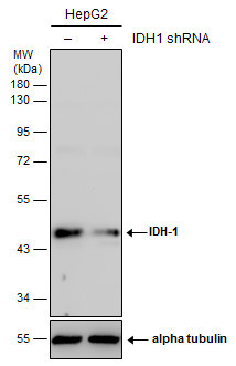
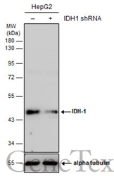
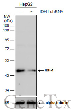
- Non-transfected (¡V) and transfected (+) HepG2 whole cell extracts (30 ?g) were separated by 10% SDS-PAGE, and the membrane was blotted with IDH1 antibody (GTX105179) diluted at 1:5000. The HRP-conjugated anti-rabbit IgG antibody (GTX213110-01) was used to detect the primary antibody.
Supportive validation
- Submitted by
- GeneTex (provider)
- Main image

- Experimental details
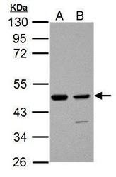
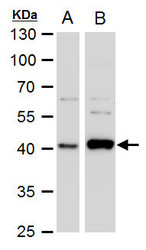
- Sample (30 ug of whole cell lysate) A: A431 B: HepG2 10% SDS PAGE GTX105179 diluted at 1:1000
- Validation comment
- WB

- Submitted by
- GeneTex (provider)
- Main image
- Experimental details
- Sample (30 ?g of whole cell lysate) A: JC B: C2C12 10% SDS PAGE GTX105179 diluted at 1:1000 The HRP-conjugated anti-rabbit IgG antibody (GTX213110-01) was used to detect the primary antibody.

- Submitted by
- GeneTex (provider)
- Main image
- Experimental details
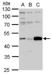
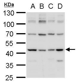
- IDH1 antibody detects IDH1 protein by western blot analysis.A. 30 ?g 293T whole cell lysate/extract B. 30 ?g HeLa whole cell lysate/extract C. 30 ?g HepG2 whole cell lysate/extract10% SDS-PAGEIDH1 antibody (GTX105179) dilution: 1:1000The HRP-conjugated anti-rabbit IgG antibody (GTX213110-01) was used to detect the primary antibody.

- Submitted by
- GeneTex (provider)
- Main image
- Experimental details
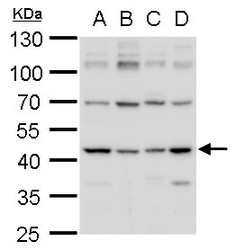
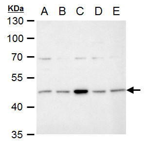
- IDH1 antibody detects IDH1 protein by western blot analysis.A. 30 ?g Neuro2A whole cell lysate/extract B. 30 ?g GL261 whole cell lysate/extract C. 30 ?g NIH-3T3 whole cell lysate/extract D. 30 ?g BCL-1 whole cell lysate/extract10% SDS-PAGEIDH1 antibody (GTX105179) dilution: 1:1000The HRP-conjugated anti-rabbit IgG antibody (GTX213110-01) was used to detect the primary antibody.
- Submitted by
- GeneTex (provider)
- Main image
- Experimental details
- Non-transfected (¡V) and transfected (+) HepG2 whole cell extracts (30 ?g) were separated by 10% SDS-PAGE, and the membrane was blotted with IDH1 antibody (GTX105179) diluted at 1:5000. The HRP-conjugated anti-rabbit IgG antibody (GTX213110-01) was used to detect the primary antibody.
- Submitted by
- GeneTex (provider)
- Main image
- Experimental details
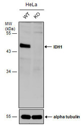
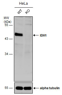
- Wild-type (WT) and IDH1 knockout (KO) HeLa cell extracts (30 ?g) were separated by 10% SDS-PAGE, and the membrane was blotted with IDH1 antibody (GTX105179) diluted at 1:1000. The HRP-conjugated anti-rabbit IgG antibody (GTX213110-01) was used to detect the primary antibody, and the signal was developed with Trident ECL plus-Enhanced.
- Submitted by
- GeneTex (provider)
- Main image
- Experimental details
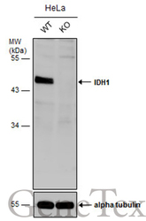
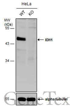
- Wild-type (WT) and IDH1 knockout (KO) HeLa cell extracts (30 ?g) were separated by 10% SDS-PAGE, and the membrane was blotted with IDH1 antibody (GTX105179) diluted at 1:1000. The HRP-conjugated anti-rabbit IgG antibody (GTX213110-01) was used to detect the primary antibody, and the signal was developed with Trident ECL plus-Enhanced.
- Submitted by
- GeneTex (provider)
- Main image
- Experimental details
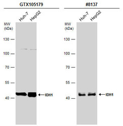
- Various whole cell extracts (30 ?g) were separated by 7.5% SDS-PAGE, and the membranes were blotted with IDH1 antibody (GTX105179) diluted at 1:1000 and competitor's antibody (CST#8137) diluted at 1:1000. The HRP-conjugated anti-rabbit IgG antibody (GTX213110-01) was used to detect the primary antibody.

- Submitted by
- GeneTex (provider)
- Main image
- Experimental details
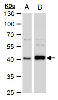
- IDH1 antibody detects IDH1 protein by Western blot analysis.A. 30 £gg A431 whole cell extract B. 30 £gg HepG2 whole cell extract 10 % SDS-PAGEIDH1 antibody (GTX105179) dilution: 1:1000
- Submitted by
- GeneTex (provider)
- Main image
- Experimental details
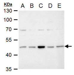
- IDH1 antibody detects IDH1 protein by Western blot analysis.A. 30 £gg Neuro2A whole cell extract B. 30 £gg C8D30 whole cell extract C. 30 £gg NIH-3T3 whole cell extract D. 30 £gg Raw 264.7 whole cell extract E. 30 £gg C2Cl2 whole cell extract10 % SDS-PAGEIDH1 antibody (GTX105179) dilution: 1:1000
Supportive validation
- Submitted by
- GeneTex (provider)
- Main image
- Experimental details
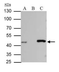
- IDH-1 antibody immunoprecipitates IDH-1 protein in IP experiments. IP Sample: HepG2 whole cell lysate/extract A : 30 £gg whole cell lysate/extract of IDH1 protein expressing HepG2 cells B : Control with 2.5 £gg of pre-immune rabbit IgG C : Immunoprecipitation of IDH-1 protein by 2.5 £gg of IDH-1 antibody (GTX105179) 10% SDS-PAGE The immunoprecipitated IDH-1 protein was detected by IDH-1 antibody (GTX105179) diluted at 1 : 1000. EasyBlot anti-rabbit IgG (HRP) (GTX221666-01) was used as a secondary reagent.

Supportive validation
- Submitted by
- GeneTex (provider)
- Main image
- Experimental details
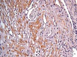
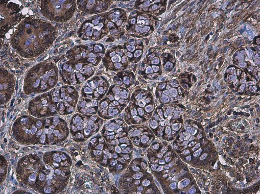
- Immunohistochemical analysis of paraffin-embedded human colon carcinoma, using IDH1(GTX105179) antibody at 1:100 dilution.
- Submitted by
- GeneTex (provider)
- Main image
- Experimental details
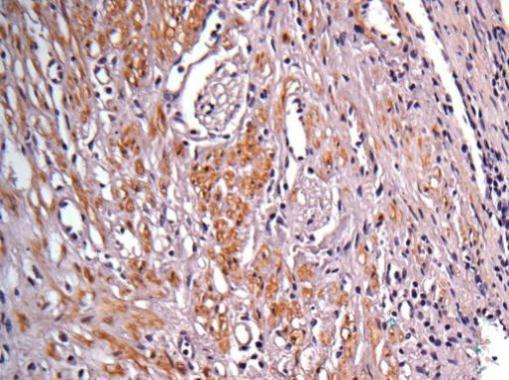
- IDH1 antibody detects IDH1 protein at cytoplasm in rat colon by immunohistochemical analysis. Sample: Paraffin-embedded rat colon. IDH1 antibody (GTX105179) diluted at 1:500.
- Submitted by
- GeneTex (provider)
- Main image
- Experimental details
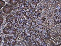
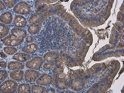
- IDH1 antibody detects IDH1 protein at cytoplasm in mouse duodenum by immunohistochemical analysis. Sample: Paraffin-embedded mouse duodenum. IDH1 antibody (GTX105179) diluted at 1:500.
