 Explore
 Validate
 Learn
Learn Western blot
Antibody data
- Antibody Data
- Antigen structure
- References [0]
- Comments [0]
- Validations
- Western blot [1]
- Immunocytochemistry [1]
- Immunoprecipitation [1]
- Immunohistochemistry [1]
Submit
Validation data
Reference
Comment
Report error
- Product number
- GTX100092 - Provider product page

- Provider
- GeneTex
- Proper citation
- GeneTex Cat#GTX100092, RRID:AB_1241458
- Product name
- XLF antibody [N3C3]
- Antibody type
- Polyclonal
- Reactivity
- Human
- Host
- Rabbit
No comments: Submit comment
Supportive validation
- Submitted by
- GeneTex (provider)
- Main image

- Experimental details
- Sample (30 ug of whole cell lysate) A: Hep G2 (GTX27900) 12% SDS PAGE GTX100092 diluted at 1:1000

Supportive validation
- Submitted by
- GeneTex (provider)
- Main image

- Experimental details
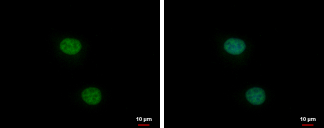
- XLF antibody [N3C3] detects XLF protein at nucleus by immunofluorescent analysis.Sample: HeLa cells were fixed in 4% paraformaldehyde at RT for 15 min.Green: XLF protein stained by XLF antibody [N3C3] (GTX100092) diluted at 1:1000.Blue: Hoechst 33342 staining.
Supportive validation
- Submitted by
- GeneTex (provider)
- Main image

- Experimental details
- XLF antibody [N3C3] immunoprecipitates XLF protein in IP experiments.IP samples: Jurkat whole cell extractA. Control with 3 £gg of preimmune Rabbit IgGB. Immunoprecipitation of XLF protein by 3 £gg XLF antibody [N3C3] (GTX100092)10 % SDS-PAGEThe immunoprecipitated XLF protein was detected by XLF antibody [N3C3] (GTX100092) diluted at 1:1000.[EasyBlot anti-rabbit IgG (GTX221666-01) was used as a secondary reagent]

Supportive validation
- Submitted by
- GeneTex (provider)
- Main image

- Experimental details
- XLF antibody [N3C3] detects XLF protein on human ovarian carcinoma by immunohistochemical analysis. Sample: Paraffin-embedded ovarian carcinoma. XLF antibody [N3C3] (GTX100092) dilution: 1:500.
