 Explore
 Validate
 Learn
Learn Western blot
Antibody data
- Antibody Data
- Antigen structure
- References [2]
- Comments [0]
- Validations
- Western blot [3]
- Other assay [1]
Submit
Validation data
Reference
Comment
Report error
- Product number
- PA1-25081 - Provider product page

- Provider
- Invitrogen Antibodies
- Product name
- Catenin alpha-1 Polyclonal Antibody
- Antibody type
- Polyclonal
- Antigen
- Synthetic peptide
- Description
- Recommended positive controls: A431 and cytosolic fraction of rat embryonic brain, MDCK, MCF-7. PA1-25081 is expected to cross react with mouse (100% conserved) and Xenopus laevis (100% conserved) due to sequence homology. Store product as a concentrated solution. Centrifuge briefly prior to opening the vial.
- Reactivity
- Human, Mouse, Rat, Canine, Xenopus
- Host
- Rabbit
- Isotype
- IgG
- Vial size
- 100 µL
- Storage
- Store at 4°C short term. For long term storage, store at -20°C, avoiding freeze/thaw cycles.
Submitted references Tight junctions negatively regulate mechanical forces applied to adherens junctions in vertebrate epithelial tissue.
Actomyosin-generated tension on cadherin is similar between dividing and non-dividing epithelial cells in early Xenopus laevis embryos.
Hatte G, Prigent C, Tassan JP
Journal of cell science 2018 Feb 5;131(3)
Journal of cell science 2018 Feb 5;131(3)
Actomyosin-generated tension on cadherin is similar between dividing and non-dividing epithelial cells in early Xenopus laevis embryos.
Herbomel G, Hatte G, Roul J, Padilla-Parra S, Tassan JP, Tramier M
Scientific reports 2017 Mar 22;7:45058
Scientific reports 2017 Mar 22;7:45058
No comments: Submit comment
Supportive validation
- Submitted by
- Invitrogen Antibodies (provider)
- Main image

- Experimental details
- Western blot of alpha Catenin in rat embryonic brain (S1) extract using an Alpha Catenin polyclonal antibody (Product # PA1-25081) at a dilution of 1:2000 and detected using an HRP anti-Rabbit IgG and ECL substrate.

- Submitted by
- Invitrogen Antibodies (provider)
- Main image
- Experimental details
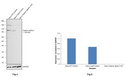
- Knockout of Catenin alpha-1 was achieved by CRISPR-Cas9 genome editing using LentiArray™ Lentiviral sgRNA (Product # A32042, Assay ID CRISPR787281_LV) and LentiArray Cas9 Lentivirus (Product # A32064). Western blot analysis of Catenin alpha-1 was performed by loading 30 µg of HeLa Wild Type (Lane 1), HeLa Cas9 (Lane 2) andHeLa Catenin alpha-1 KO (Lane 3) whole cell extracts. The samples were electrophoresed using NuPAGE™ Novex™ 4-12% Bis-Tris Protein Gel (Product # NP0322BOX). Resolved proteins were then transferred onto a nitrocellulose membrane (Product # IB23001) by iBlot® 2 Dry Blotting System (Product # IB21001). The blot was probed with Anti-Catenin alpha-1 Polyclonal Antibody (Product # PA1-25081, 1:1,000 dilution) and Goat anti-Rabbit IgG (H+L) Superclonal™ Recombinant Secondary Antibody, HRP (Product # A27036, 1:8,000 dilution) using the iBright FL 1000 (Product # A32752). Chemiluminescent detection was performed using Novex® ECL Chemiluminescent Substrate Reagent Kit (Product # WP20005). Loss of signal upon CRISPR mediated knockout (KO) using the LentiArray™ CRISPR product line confirms that antibody is specific to Catenin alpha-1. An uncharacterized band was observed in all the samples at ~160 kDa.

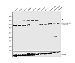
- Submitted by
- Invitrogen Antibodies (provider)
- Main image
- Experimental details
- Western blot was performed using Anti-Catenin alpha-1 Polyclonal Antibody(Product # PA1-25081) and a 100kDa band corresponding to Catenin alpha-1 was observed across cell lines and tissue extracts tested except PC-3 and Daudi which is reported to be negative. A non-specific band around 160kDA has also been observed in cell lines. Whole Cell Extract-WCL (30 µg lysate) of A-431 (Lane 1), HeLa (Lane 2), MCF7 (Lane 3), LNCaP (Lane 4), PC-3 (Lane 5), Daudi (Lane 6), NIH/3T3 (Lane 7), PC-12 (Lane 8) and tissue extracts of Mouse Lung (Lane 9), Mouse Heart (Lane 10) and Rat Heart (Lane 11) were electrophoresed using NuPAGE™ 4-12% Bis-Tris Protein Gel (Product # NP0322BOX). Resolved proteins were then transferred onto a Nitrocellulose membrane (Product # IB23001) by iBlot® 2 Dry Blotting System (Product # IB21001). The blot was probed with the primary antibody (1:1000 dilution) and detected by chemiluminescence with Goat anti-Rabbit IgG (H+L) Superclonal™ Recombinant Secondary Antibody, HRP (Product # A27036, 1:4000 dilution) using the iBright FL 1000 (Product # A32752). Chemiluminescent detection was performed using Novex® ECL Chemiluminescent Substrate Reagent Kit (Product # WP20005).

Supportive validation
- Submitted by
- Invitrogen Antibodies (provider)
- Main image

- Experimental details
- Figure 3 EcadTSMod biosensor is integrated into the adherens junction and responds to several treatments. ( a ) Scatter plot of the fluorescence lifetime vs fluorescence intensity for each pixel of a representative acquisition. Equation of the linear regression is specified in the top right corner. ( b ) Indirect immunofluorescences of EcadTSMod detected with anti-GFP antibody (green) and alpha, beta and p120 catenins (red) in Xenopus laevis blastula overexpressing the EcadTSMod protein. Linescan were made on the indicated dotted line, and the normalized fluorescence intensity is shown for both channel (green for EcadTSMod and red for catenins) in the corresponding graph. Scale bar, 20 mum. ( c ) Boxplot of mean forces applied on EcadTSMod in untreated embryos or embryos treated with EGTA, latrunculin A, morpholino against alpha-catenin (MO alpha-catenin) and Calyculin A, with respectively 105, 32, 37, 44 and 29 independent fields (respective number of experiments: 14, 2, 2, 2 and 3; respective number of embryos: 55, 16, 19, 22 and 15). For each comparison, the p value is indicated. In the box plot, bold bars correspond to median value, whiskers to the value 1.5x away from the 1 st and 3 rd quartiles, and the dot to outlier.
