 Explore
 Validate
 Learn
Learn Western blot
Antibody data
- Antibody Data
- Antigen structure
- References [2]
- Comments [0]
- Validations
- Western blot [3]
- Immunocytochemistry [2]
- Immunohistochemistry [2]
Submit
Validation data
Reference
Comment
Report error
- Product number
- GTX111095 - Provider product page

- Provider
- GeneTex
- Proper citation
- GeneTex Cat#GTX111095, RRID:AB_1950058
- Product name
- alpha 1 Catenin antibody [N3C2], Internal
- Antibody type
- Polyclonal
- Reactivity
- Human, Mouse, Rat, Zebrafish
- Host
- Rabbit
Submitted references Nuclear PGK1 Alleviates ADP-Dependent Inhibition of CDC7 to Promote DNA Replication.
Sequential and opposing activities of Wnt and BMP coordinate zebrafish bone regeneration.
Li X, Qian X, Jiang H, Xia Y, Zheng Y, Li J, Huang BJ, Fang J, Qian CN, Jiang T, Zeng YX, Lu Z
Molecular cell 2018 Nov 15;72(4):650-660.e8
Molecular cell 2018 Nov 15;72(4):650-660.e8
Sequential and opposing activities of Wnt and BMP coordinate zebrafish bone regeneration.
Stewart S, Gomez AW, Armstrong BE, Henner A, Stankunas K
Cell reports 2014 Feb 13;6(3):482-98
Cell reports 2014 Feb 13;6(3):482-98
No comments: Submit comment
Supportive validation
- Submitted by
- GeneTex (provider)
- Main image

- Experimental details
- Sample (30 ug of whole cell lysate) A: NIH-3T3 B: JC 7.5% SDS PAGE GTX111095 diluted at 1:10000

- Submitted by
- GeneTex (provider)
- Main image

- Experimental details
- Sample (30 ug of whole cell lysate) A: PC-12 7.5% SDS PAGE GTX111095 diluted at 1:10000

- Submitted by
- GeneTex (provider)
- Main image

- Experimental details
- Sample (30 ug of whole cell lysate) A: Hep G2 (GTX27900) 7.5% SDS PAGE GTX111095 diluted at 1:1000

Supportive validation
- Submitted by
- GeneTex (provider)
- Main image

- Experimental details
- alpha 1 Catenin antibody [N3C2], Internal detects CTNNA1 protein at junction by immunofluorescent analysis. Sample: HeLa cells were fixed in 4% paraformaldehyde at RT for 15 min.Green: CTNNA1 protein stained by alpha 1 Catenin antibody [N3C2], Internal (GTX111095) diluted at 1:500.Blue: Hoechst 33342 staining.

- Submitted by
- GeneTex (provider)
- Main image

- Experimental details
- alpha 1 catenin antibody [N3c2], Internal detects alpha 1 Catenin protein at cytoplasm and cell junction by immunofluorescent analysis.Sample: HeLa cells were fixed in 4% paraformaldehyde at RT for 15 min.Green: alpha 1 Catenin protein stained by alpha 1 catenin antibody [N3c2], Internal (GTX111095) diluted at 1:500.Red: alpha Tubulin, a cytoskeleton marker, stained by alpha Tubulin antibody [GT114] (GTX628802) diluted at 1:500.Blue: Hoechst 33342 staining.

Supportive validation
- Submitted by
- GeneTex (provider)
- Main image
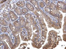
- Experimental details
- alpha 1 Catenin antibody [N3C2], Internal detects alpha 1 Catenin protein at membrane on mouse intestine by immunohistochemical analysis. Sample: Paraffin-embedded mouse intestine. alpha 1 Catenin antibody [N3C2], Internal (GTX111095) dilution: 1:500.

- Submitted by
- GeneTex (provider)
- Main image

- Experimental details
- alpha 1 Catenin antibody [N3C2], Internal detects alpha 1 Catenin protein at membrane on mouse prostate by immunohistochemical analysis. Sample: Paraffin-embedded mouse prostate. alpha 1 Catenin antibody [N3C2], Internal (GTX111095) dilution: 1:500.
