Explore
 Validate
 Learn
Learn Western blot
 Flow cytometry
Antibody data
- Antibody Data
- Antigen structure
- References [12]
- Comments [0]
- Validations
- Western blot [3]
- Immunocytochemistry [1]
- Immunohistochemistry [2]
Submit
Validation data
Reference
Comment
Report error
- Product number
- MA1-06318 - Provider product page

- Provider
- Invitrogen Antibodies
- Product name
- Cytokeratin 8 Monoclonal Antibody (M20)
- Antibody type
- Monoclonal
- Antigen
- Other
- Description
- MA1-06318 detects cytokeratin 8 in rat, human, and rabbit samples. MA1-06318 has sucessfully been used in Western blotting, immunocytochemistry, flow cytometry and immunohistochemistry (frozen, paraffin). The MA1-06318 immunogen is keratin isolated from the human breast carcinoma cell line MCF-7. Store at 4ºC or in small aliquots at -20ºC.
- Reactivity
- Human, Rat, Rabbit
- Host
- Mouse
- Isotype
- IgG
- Antibody clone number
- M20
- Vial size
- 100 µL
- Concentration
- 1 mg/mL
- Storage
- Store at 4°C short term. For long term storage, store at -20°C, avoiding freeze/thaw cycles.
Submitted references Keratin 8 phosphorylation regulates its transamidation and hepatocyte Mallory-Denk body formation.
Generation of hepatocyte-like cells from in vitro transdifferentiated human fetal pancreas.
Generation of hepatocyte-like cells from in vitro transdifferentiated human fetal pancreas.
Antigen expression of human eccrine sweat glands.
Analysis of keratin polypeptides 8 and 19 variants in inflammatory bowel disease.
Bispecific and human disease-related anti-keratin rabbit monoclonal antibodies.
Protein phosphatase-2A associates with and dephosphorylates keratin 8 after hyposmotic stress in a site- and cell-specific manner.
Mesenchymal hamartoma of the liver in adulthood: immunohistochemical profiles, clinical and histopathological features in two patients.
Studying simple epithelial keratins in cells and tissues.
Keratin 8 and 18 hyperphosphorylation is a marker of progression of human liver disease.
Type II keratins are phosphorylated on a unique motif during stress and mitosis in tissues and cultured cells.
Keratin 8 mutations in patients with cryptogenic liver disease.
Kwan R, Hanada S, Harada M, Strnad P, Li DH, Omary MB
FASEB journal : official publication of the Federation of American Societies for Experimental Biology 2012 Jun;26(6):2318-26
FASEB journal : official publication of the Federation of American Societies for Experimental Biology 2012 Jun;26(6):2318-26
Generation of hepatocyte-like cells from in vitro transdifferentiated human fetal pancreas.
Sumitran-Holgersson S, Nowak G, Thowfeequ S, Begum S, Joshi M, Jaksch M, Kjaeldgaard A, Jorns C, Ericzon BG, Tosh D
Cell transplantation 2009;18(2):183-93
Cell transplantation 2009;18(2):183-93
Generation of hepatocyte-like cells from in vitro transdifferentiated human fetal pancreas.
Sumitran-Holgersson S, Nowak G, Thowfeequ S, Begum S, Joshi M, Jaksch M, Kjaeldgaard A, Jorns C, Ericzon BG, Tosh D
Cell transplantation 2009;18(2):183-93
Cell transplantation 2009;18(2):183-93
Antigen expression of human eccrine sweat glands.
Li HH, Zhou G, Fu XB, Zhang L
Journal of cutaneous pathology 2009 Mar;36(3):318-24
Journal of cutaneous pathology 2009 Mar;36(3):318-24
Analysis of keratin polypeptides 8 and 19 variants in inflammatory bowel disease.
Tao GZ, Strnad P, Zhou Q, Kamal A, Zhang L, Madani ND, Kugathasan S, Brant SR, Cho JH, Omary MB, Duerr RH
Clinical gastroenterology and hepatology : the official clinical practice journal of the American Gastroenterological Association 2007 Jul;5(7):857-64
Clinical gastroenterology and hepatology : the official clinical practice journal of the American Gastroenterological Association 2007 Jul;5(7):857-64
Bispecific and human disease-related anti-keratin rabbit monoclonal antibodies.
Tao GZ, Nakamichi I, Ku NO, Wang J, Frolkis M, Gong X, Zhu W, Pytela R, Omary MB
Experimental cell research 2006 Feb 15;312(4):411-22
Experimental cell research 2006 Feb 15;312(4):411-22
Protein phosphatase-2A associates with and dephosphorylates keratin 8 after hyposmotic stress in a site- and cell-specific manner.
Tao GZ, Toivola DM, Zhou Q, Strnad P, Xu B, Michie SA, Omary MB
Journal of cell science 2006 Apr 1;119(Pt 7):1425-32
Journal of cell science 2006 Apr 1;119(Pt 7):1425-32
Mesenchymal hamartoma of the liver in adulthood: immunohistochemical profiles, clinical and histopathological features in two patients.
Yesim G, Gupse T, Zafer U, Ahmet A
Journal of hepato-biliary-pancreatic surgery 2005;12(6):502-7
Journal of hepato-biliary-pancreatic surgery 2005;12(6):502-7
Studying simple epithelial keratins in cells and tissues.
Ku NO, Toivola DM, Zhou Q, Tao GZ, Zhong B, Omary MB
Methods in cell biology 2004;78:489-517
Methods in cell biology 2004;78:489-517
Keratin 8 and 18 hyperphosphorylation is a marker of progression of human liver disease.
Toivola DM, Ku NO, Resurreccion EZ, Nelson DR, Wright TL, Omary MB
Hepatology (Baltimore, Md.) 2004 Aug;40(2):459-66
Hepatology (Baltimore, Md.) 2004 Aug;40(2):459-66
Type II keratins are phosphorylated on a unique motif during stress and mitosis in tissues and cultured cells.
Toivola DM, Zhou Q, English LS, Omary MB
Molecular biology of the cell 2002 Jun;13(6):1857-70
Molecular biology of the cell 2002 Jun;13(6):1857-70
Keratin 8 mutations in patients with cryptogenic liver disease.
Ku NO, Gish R, Wright TL, Omary MB
The New England journal of medicine 2001 May 24;344(21):1580-7
The New England journal of medicine 2001 May 24;344(21):1580-7
No comments: Submit comment
Supportive validation
- Submitted by
- Invitrogen Antibodies (provider)
- Main image

- Experimental details
- CRISPR-Cas9 mediated genome editing ofCytokeratin 8 (as confirmed by next generation sequencing) was achieved by using LentiArray™ Lentiviral sgRNA (Product # A32042, AssayID CRISPR745510_LV) and LentiArray Cas9 Lentivirus (Product # A32064). Fig (a) Western blot analysis of Cytokeratin 8 was performed by loading 30 µg of HeLa Wild Type (Lane 1), HeLa Cas9 (Lane 2) and HeLa Cas9 cells transduced with Cytokeratin 8 Lentiviral sgRNA (Lane 3) whole cell extracts. The samples were electrophoresed using NuPAGE™ Novex™ 4-12% Bis-Tris Protein Gel (Product # NP0322BOX). Resolved proteins were then transferred onto a nitrocellulose membrane (Product # IB23001) by iBlot® 2 Dry Blotting System (Product # IB21001). The blot was probed with Anti-Cytokeratin 8 Monoclonal Antibody (M20) (Product # MA1-06318) using 1:1,000 dilution and Goat anti-Mouse IgG (H+L) Superclonal™ Recombinant Secondary Antibody, HRP (Product # A28177 1:5,000 dilution).Chemiluminescent detection was performed using Novex® ECL Chemiluminescent Substrate Reagent Kit (Product # WP20005). A loss of signal in sgRNA transduced cells using the LentiArray™ CRISPR product line confirms that antibody is specific toCytokeratin 8 (Fig (b)).

- Submitted by
- Invitrogen Antibodies (provider)
- Main image

- Experimental details
- Knockdown of Cytokeratin 8 was achieved by transfecting Caco-2 with Cytokeratin 8 specific siRNAs (Silencer® select Product # S7969, S7970). Western blot analysis (Fig. a) was performed using Whole cell extracts from the Cytokeratin 8 knockdown cells (lane 3), non-targeting scrambled siRNA transfected cells (lane 2) and untransfected cells (lane 1). The blot was probed with Cytokeratin 8 Monoclonal Antibody (M20) (Product # MA1-06318, 1:1000 dilution ) and Goat anti-Mouse IgG (H+L) Superclonal™ Recombinant Secondary Antibody, HRP (Product # A28177, 1:4000 dilution). Densitometric analysis of this western blot is shown in histogram (Fig. b). Decrease in signal upon siRNA mediated knock down confirms that antibody is specific to Cytokeratin 8.

- Submitted by
- Invitrogen Antibodies (provider)
- Main image
- Experimental details
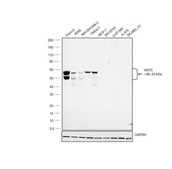
- Western blot was performed using Anti-Cytokeratin 8 Monoclonal Antibody (M20)(Product # MA1-06318) and a 56 & 53 kDa band corresponding to Cytokeratin 8 was observed across the cell lines tested except SH-SY5Y, U-87 MG, A-375 and SK-MEL-31. Whole cell extracts (30 µg lysate) of Caco-2 (Lane 1), A549 (Lane 2), NIH:OVCAR-3 (Lane 3), HaCaT (Lane 4), MCF7 (Lane 5), SH-SY5Y (Lane 6), U-87 MG (Lane 7), A-375 (Lane 8) and SK-MEL-31(Lane 9) were electrophoresed using NuPAGE™ 4-12% Bis-Tris Protein Gel (Product # NP0321BOX). Resolved proteins were then transferred onto a Nitrocellulose membrane (Product # LC2001) by iBlot® 2 Dry Blotting System (Product # IB21001). The blot was probed with the primary antibody (1:1000 dilution) and detected by chemiluminescence with Goat anti-Mouse IgG (H+L) Superclonal™ Recombinant Secondary Antibody, HRP (Product # A28177, 1:4000 dilution) using the iBright FL 1000 (Product # A32752). Chemiluminescent detection was performed using Novex® ECL Chemiluminescent Substrate Reagent Kit (Product # WP20005).

Supportive validation
- Submitted by
- Invitrogen Antibodies (provider)
- Main image

- Experimental details
- Immunofluorescence analysis of Cytokeratin 8 was performed using 70% confluent log phase Caco-2 cells. The cells were fixed with ice-cold acetone at 4°C for 5 minutes and blocked with 2% BSA for 45 minutes at room temperature. The cells were labeled with Cytokeratin 8 Monoclonal Antibody (M20) (Product # MA1-06318) at 1:200 dilution in 0.1% BSA, incubated at 4 degree celsius overnight and then labeled with Goat anti-Mouse IgG (H+L) Superclonal™ Recombinant Secondary Antibody, Alexa Fluor® 488 conjugate (Product # A28175), (1:2000 dilution), for 45 minutes at room temperature (Panel a: Green). Nuclei (Panel b:Blue) were stained with SlowFade® Gold Antifade Mountant with DAPI (Product # S36938). F-actin (Panel c: Red) was stained with Rhodamine Phalloidin (Product # R415, 1:300 dilution). Panel d represents the merged image showing cytoskeletal localization. Panel e represents control cells with no primary antibody to assess background. The images were captured at 60X magnification.

Supportive validation
- Submitted by
- Invitrogen Antibodies (provider)
- Main image

- Experimental details
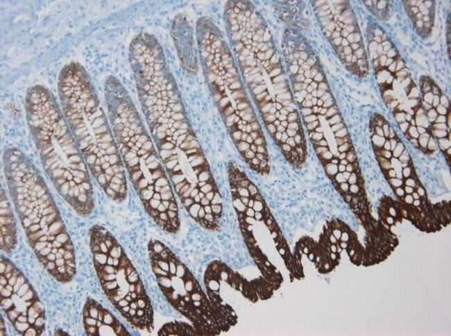
- Immunohistochemistry on frozen section of human colon stained with Cytokeratin 8 monoclonal antibody (Product # MA1-06318).

- Submitted by
- Invitrogen Antibodies (provider)
- Main image

- Experimental details
- Immunohistochemistry on paraffin section of human colon stained with Cytokeratin 8 monoclonal antibody (Product # MA1-06318).