Explore
 Validate
 Learn
Learn Western blot
Antibody data
- Antibody Data
- Antigen structure
- References [0]
- Comments [0]
- Validations
- Western blot [6]
- Immunocytochemistry [2]
- Immunohistochemistry [1]
Submit
Validation data
Reference
Comment
Report error
- Product number
- PA5-34762 - Provider product page

- Provider
- Invitrogen Antibodies
- Product name
- Cytokeratin 8 Polyclonal Antibody
- Antibody type
- Polyclonal
- Antigen
- Recombinant protein fragment
- Description
- Recommended positive controls: HeLa, HepG2, JC. Predicted reactivity: Mouse (86%), Rat (85%), Dog (88%), Pig (82%), Rhesus Monkey (99%), Bovine (84%). Store product as a concentrated solution. Centrifuge briefly prior to opening the vial.
- Reactivity
- Human, Mouse
- Host
- Rabbit
- Isotype
- IgG
- Vial size
- 100 µL
- Concentration
- 0.66 mg/mL
- Storage
- Store at 4°C short term. For long term storage, store at -20°C, avoiding freeze/thaw cycles.
No comments: Submit comment
Supportive validation
- Submitted by
- Invitrogen Antibodies (provider)
- Main image
- Experimental details
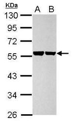
- Western blot analysis of Cytokeratin 8 using 30 µg of A) HeLa and B) HepG2 lysate. Samples were loaded onto a 10% SDS-PAGE gel and probed with a Cytokeratin 8 polyclonal antibody (Product # PA5-34762) at a dilution of 1:10,000.

- Submitted by
- Invitrogen Antibodies (provider)
- Main image

- Experimental details
- CRISPR-Cas9 mediated genome editing ofCytokeratin 8 (as confirmed by next generation sequencing) was achieved by using LentiArray™ Lentiviral sgRNA (Product # A32042, AssayID CRISPR745510_LV) and LentiArray Cas9 Lentivirus (Product # A32064). Fig (a) Western blot analysis of Cytokeratin 8 was performed by loading 30 µg of HeLa Wild Type (Lane 1), HeLa Cas9 (Lane 2) and HeLa Cas9 cells transduced with Cytokeratin 8 Lentiviral sgRNA (Lane 3) whole cell extracts. The samples were electrophoresed using NuPAGE™ Novex™ 4-12% Bis-Tris Protein Gel (Product # NP0322BOX). Resolved proteins were then transferred onto a nitrocellulose membrane (Product # IB23001) by iBlot® 2 Dry Blotting System (Product # IB21001). The blot was probed with Anti-Cytokeratin 8 Polyclonal Antibody (Product # PA5-34762) using 1:10,000 dilution and Goat anti-Rabbit IgG (H+L) Superclonal™ Recombinant Secondary Antibody, HRP (Product # A27036 1:5,000 dilution).Chemiluminescent detection was performed using Novex® ECL Chemiluminescent Substrate Reagent Kit (Product # WP20005). A loss of signal in sgRNA transduced cells using the LentiArray™ CRISPR product line confirms that antibody is specific toCytokeratin 8 (Fig (b)).

- Submitted by
- Invitrogen Antibodies (provider)
- Main image

- Experimental details
- Western Blot using Cytokeratin 8 Polyclonal Antibody (Product # PA5-34762). Sample (30 µg of whole cell lysate). Lane A: JC. 10% SDS PAGE. Cytokeratin 8 Polyclonal Antibody (Product # PA5-34762) diluted at 1:10,000. The HRP-conjugated anti-rabbit IgG antibody was used to detect the primary antibody.

- Submitted by
- Invitrogen Antibodies (provider)
- Main image

- Experimental details
- Western Blot using Cytokeratin 8 Polyclonal Antibody (Product # PA5-34762). Various whole cell extracts (30 µg) were separated by 10% SDS-PAGE, and the membrane was blotted with Cytokeratin 8 Polyclonal Antibody (Product # PA5-34762) diluted at 1:10,000. The HRP-conjugated anti-rabbit IgG antibody was used to detect the primary antibody.

- Submitted by
- Invitrogen Antibodies (provider)
- Main image
- Experimental details
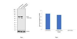
- Knockdown of Cytokeratin 8 was achieved by transfecting Caco-2 with Cytokeratin 8 specific siRNAs (Silencer® select Product # S7969, S7970). Western blot analysis (Fig. a) was performed using Whole cell extracts from the Cytokeratin 8 knockdown cells (lane 3), non-targeting scrambled siRNA transfected cells (lane 2) and untransfected cells (lane 1). The blot was probed with Cytokeratin 8 Polyclonal Antibody (Product # PA5-34762,1:10000 dilution) and Goat anti-Rabbit IgG (H+L) Superclonal™ Recombinant Secondary Antibody, HRP (Product # A27036, 1:4000 dilution). Densitometric analysis of this western blot is shown in histogram (Fig. b). Decrease in signal upon siRNA mediated knock down confirms that antibody is specific to Cytokeratin 8.

- Submitted by
- Invitrogen Antibodies (provider)
- Main image

- Experimental details
- Western blot was performed using Anti-Cytokeratin 8 Polyclonal Antibody(Product # PA5-34762) and 53, 57kDa bands corresponding to Cytokeratin 8 was observed across the cell lines tested except IMR-32, SH-SY5Y and SK-MEL-31. Whole cell extracts (30 µg lysate) of Caco-2 (Lane 1), HaCaT (Lane 2), MCF-7 (Lane 3), IMR-32 (Lane 4), SH-SY5Y (Lane 5), and SK-MEL-31 (Lane 6) were electrophoresed using NuPAGE™ 4-12% Bis-Tris Protein Gel (Product # NP0321BOX). Resolved proteins were then transferred onto a Nitrocellulose membrane (Product # LC2002) by iBlot® 2 Dry Blotting System (Product # IB21001). The blot was probed with the primary antibody (1:10000 dilution) and detected by chemiluminescence with Goat anti-Rabbit IgG (H+L) Superclonal™ Recombinant Secondary Antibody, HRP (Product # A27036,1:4000 dilution) using the iBright FL 1000 (Product # A32752). Chemiluminescent detection was performed using Novex® ECL Chemiluminescent Substrate Reagent Kit (Product # WP20005).

Supportive validation
- Submitted by
- Invitrogen Antibodies (provider)
- Main image

- Experimental details
- Immunocytochemistry-Immunofluorescence analysis of Cytokeratin 8 was performed in HeLa cells fixed in 4% paraformaldehyde at RT for 15 min. Green: Cytokeratin 8 Polyclonal Antibody (Product # PA5 34762) diluted at 1:500. Blue: Hoechst 33342 staining.

- Submitted by
- Invitrogen Antibodies (provider)
- Main image

- Experimental details
- Immunofluorescence analysis of Cytokeratin 8 was performed using 70% confluent log phase Caco-2 cells. The cells were fixed with ice-cold acetone at 4°C for 5 minutes and blocked with 2% BSA for 45 minutes at room temperature. The cells were labeled with Cytokeratin 8 Polyclonal Antibody (Product # PA5-34762) at 1:200 dilution in 0.1% BSA, incubated at 4 degree celsius overnight and then labeled with Goat anti-Mouse IgG (H+L) Superclonal™ Recombinant Secondary Antibody, Alexa Fluor® 488 conjugate (Product # A28175), (1:2000 dilution), for 45 minutes at room temperature (Panel a: Green). Nuclei (Panel b:Blue) were stained with ProLong™ Diamond Antifade Mountant with DAPI (Product # P36962). F-actin (Panel c: Red) was stained with Rhodamine Phalloidin (Product # R415, 1:300 dilution). Panel d represents the merged image showing cytoskeletal localization. Panel e represents control cells with no primary antibody to assess background. The images were captured at 60X magnification.

Supportive validation
- Submitted by
- Invitrogen Antibodies (provider)
- Main image
- Experimental details
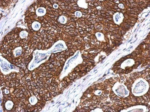
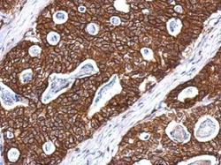
- Immunohistochemical analysis of paraffin-embedded Gastric ca, using Cytokeratin 8 (Product # PA5-34762) antibody at 1:500 dilution. Antigen Retrieval: EDTA based buffer, pH 8.0, 15 min.