 Explore
 Validate
 Learn
Learn Western blot
 Immunocytochemistry
Antibody data
- Antibody Data
- Antigen structure
- References [0]
- Comments [0]
- Validations
- Western blot [3]
- Immunohistochemistry [1]
- Flow cytometry [1]
Submit
Validation data
Reference
Comment
Report error
- Product number
- NBP2-34265-0.1mg - Provider product page

- Provider
- Novus Biologicals
- Product name
- Mouse Monoclonal Cytokeratin 8 Antibody
- Antibody type
- Monoclonal
- Description
- Protein G purified. Cytokeratin 8 (CK8) belongs to the type II (or B or basic) subfamily of high molecular weight cytokeratins and exists in combination with cytokeratin 18 (CK18). CK8 is primarily found in the non-squamous epithelia and is present in majority of adenocarcinomas and ductal carcinomas. It is absent in squamous cell carcinomas. Hepatocellular carcinomas are defined by the use of antibodies that recognize only cytokeratin 8 and 18. CK8 exists on several types of normal and neoplastic epithelia, including many ductal and glandular epithelia such as colon, stomach, small intestine, trachea, and esophagus as well as in transitional epithelium. Anti-CK8 does not react with skeletal muscle or nerve cells. Epithelioid sarcoma, chordoma, and adamantinoma show strong positivity corresponding to that of simple epithelia (with antibodies against CK8, CK18 and CK19). Reportedly, anti-CK8 is useful for the differentiation of lobular ( ring-like, perinuclear ) from ductal ( peripheral-predominant ) carcinoma of the breast.
- Reactivity
- Human, Rat, Zebrafish
- Host
- Mouse
- Isotype
- IgG
- Vial size
- 0.1 mg
- Concentration
- 0.2 mg/ml
- Storage
- Store at 4C.
No comments: Submit comment
Supportive validation
- Submitted by
- Novus Biologicals (provider)
- Main image
- Experimental details
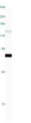
- Simple Western: Cytokeratin 8 Antibody (H1) [NBP2-34265] - Simple Western lane view shows a specific band for Cytokeratin 8 in 0.2 mg/ml of MCF-7 lysate(s). This experiment was performed under reducing conditions using the 12-230 kDa separation system.

- Submitted by
- Novus Biologicals (provider)
- Main image
- Experimental details
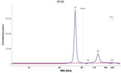
- Simple Western: Cytokeratin 8 Antibody (H1) [NBP2-34265] - Electropherogram image of the corresponding Simple Western lane. Cytokeratin 8 antibody was used at 10 ug/ml dilution of MCF-7 lysates(s) respectively.

- Submitted by
- Novus Biologicals (provider)
- Main image
- Experimental details
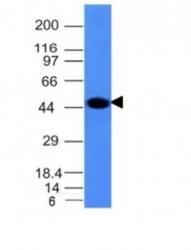
- Western Blot: Cytokeratin 8 Antibody (H1) [NBP2-34265] - Western Blot Analysis of A431 Cell Lysate using Cytokeratin 8 Monoclonal Antibody (H1)

Supportive validation
- Submitted by
- Novus Biologicals (provider)
- Main image

- Experimental details
- Immunohistochemistry-Paraffin: Cytokeratin 8 Antibody (H1) [NBP2-34265] - Formalin-fixed, paraffin-embedded human colon carcinoma stained with Cytokeratin 8 MAb (H1).

Supportive validation
- Submitted by
- Novus Biologicals (provider)
- Main image
- Experimental details
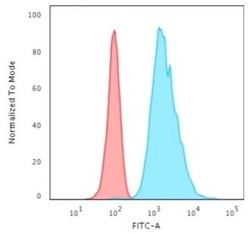
- Flow Cytometry: Cytokeratin 8 Antibody (H1) [NBP2-34265] - Flow Cytometric Analysis of HeLa cells using Cytokeratin 8 Antibody (H1) followed by Goat anti-Mouse IgG-CF488 (Blue); Isotype Control (Red).
