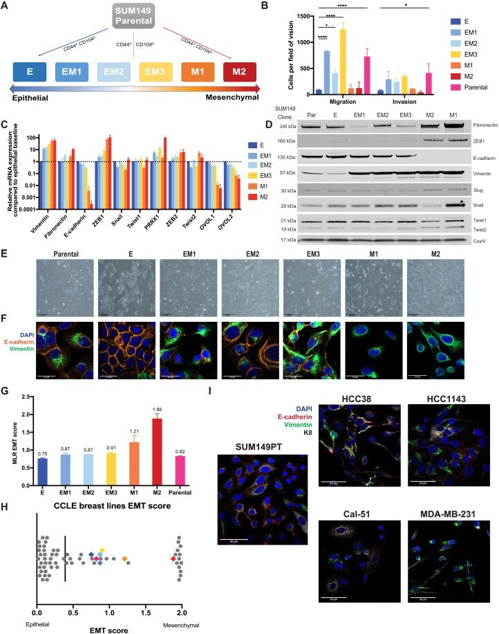
Explore
 Validate
 Learn
Learn Western blot
Antibody data
- Antibody Data
- Antigen structure
- References [3]
- Comments [0]
- Validations
- Western blot [5]
- Immunocytochemistry [1]
- Immunohistochemistry [1]
- Other assay [2]
Submit
Validation data
Reference
Comment
Report error
- Product number
- PA5-29607 - Provider product page

- Provider
- Invitrogen Antibodies
- Product name
- Cytokeratin 8 Polyclonal Antibody
- Antibody type
- Polyclonal
- Antigen
- Recombinant protein fragment
- Description
- Recommended positive controls: HepG2, HeLa, mouse liver, rat liver.
- Concentration
- 0.12 mg/mL
Submitted references Phenotypic heterogeneity driven by plasticity of the intermediate EMT state governs disease progression and metastasis in breast cancer.
Limiting Self-Renewal of the Basal Compartment by PKA Activation Induces Differentiation and Alters the Evolution of Mammary Tumors.
Skin Mucus of Gilthead Sea Bream (Sparus aurata L.). Protein Mapping and Regulation in Chronically Stressed Fish.
Brown MS, Abdollahi B, Wilkins OM, Lu H, Chakraborty P, Ognjenovic NB, Muller KE, Jolly MK, Christensen BC, Hassanpour S, Pattabiraman DR
Science advances 2022 Aug 5;8(31):eabj8002
Science advances 2022 Aug 5;8(31):eabj8002
Limiting Self-Renewal of the Basal Compartment by PKA Activation Induces Differentiation and Alters the Evolution of Mammary Tumors.
Ognjenovic NB, Bagheri M, Mohamed GA, Xu K, Chen Y, Mohamed Saleem MA, Brown MS, Nagaraj SH, Muller KE, Gerber SA, Christensen BC, Pattabiraman DR
Developmental cell 2020 Dec 7;55(5):544-557.e6
Developmental cell 2020 Dec 7;55(5):544-557.e6
Skin Mucus of Gilthead Sea Bream (Sparus aurata L.). Protein Mapping and Regulation in Chronically Stressed Fish.
Pérez-Sánchez J, Terova G, Simó-Mirabet P, Rimoldi S, Folkedal O, Calduch-Giner JA, Olsen RE, Sitjà-Bobadilla A
Frontiers in physiology 2017;8:34
Frontiers in physiology 2017;8:34
No comments: Submit comment
Supportive validation
- Submitted by
- Invitrogen Antibodies (provider)
- Main image
- Experimental details
- Western blot analysis of Cytokeratin 8 using 30 µg H1299 whole cell lysate. Samples were loaded onto a 10% SDS-PAGE gel and probed with a Cytokeratin 8 polyclonal antibody (Product # PA5-29607) at a dilution of 1:1000.
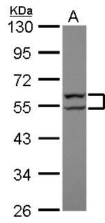
- Submitted by
- Invitrogen Antibodies (provider)
- Main image
- Experimental details
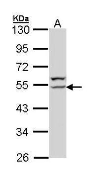
- Western blot analysis of Cytokeratin 8 using 30 µg of H1299 lysate. Samples were loaded onto a 10% SDS-PAGE gel and probed with a Cytokeratin 8 polyclonal antibody (Product # PA5-29607) at a dilution of 1:1000.
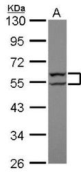
- Submitted by
- Invitrogen Antibodies (provider)
- Main image

- Experimental details
- Western blot analysis of Cytokeratin 8 was performed by separating 50 µg of rat tissue extract by 10% SDS-PAGE. Proteins were transferred to a membrane and probed with a Cytokeratin 8 Polyclonal Antibody (Product # PA5-29607) at a dilution of 1:500. The HRP-conjugated anti-rabbit IgG antibody was used to detect the primary antibody.

- Submitted by
- Invitrogen Antibodies (provider)
- Main image

- Experimental details
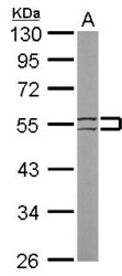
- Western Blot analysis of Cytokeratin 8 was performed by separating 30 µg of various whole cell extracts by 10% SDS-PAGE. Proteins were transferred to a membrane and probed with a Cytokeratin 8 Polyclonal Antibody (Product # PA5-29607) at a dilution of 1:2000 and a HRP-conjugated anti-rabbit IgG secondary antibody.

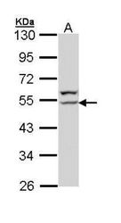
- Submitted by
- Invitrogen Antibodies (provider)
- Main image
- Experimental details
- Western Blot using Cytokeratin 8 Polyclonal Antibody (Product # PA5-29607). Sample (50 µg of whole cell lysate). Lane A: mouse liver. 10% SDS PAGE. Cytokeratin 8 Polyclonal Antibody (Product # PA5-29607) diluted at 1:1,000. The HRP-conjugated anti-rabbit IgG antibody was used to detect the primary antibody.

Supportive validation
- Submitted by
- Invitrogen Antibodies (provider)
- Main image

- Experimental details
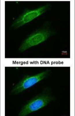
- Immunofluorescent analysis of Cytokeratin 8 in paraformaldehyde-fixed HeLa cells using a Cytokeratin 8 polyclonal antibody (Product # PA5-29607) at a 1:200 dilution.
Supportive validation
- Submitted by
- Invitrogen Antibodies (provider)
- Main image
- Experimental details
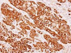
- Immunohistochemical analysis of paraffin-embedded SW480 xenograft, using Cytokeratin 8 (Product # PA5-29607) antibody at 1:500 dilution. Antigen Retrieval: EDTA based buffer, pH 8.0, 15 min.

Supportive validation
- Submitted by
- Invitrogen Antibodies (provider)
- Main image
- Experimental details
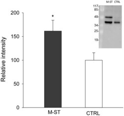
- Figure 2 Relative keratin type II cytoskeletal 8 protein levels in skin mucus of multiple sensorial stressed fish (M-ST) and control unstressed fish (CTRL) . Values of expression relative to control are the mean +- SEM of eight individuals. Asterisk indicated significant differences ( p < 0.05, Student's t -test) between groups. Insert shows a representative western blot using the rabbit anti-human cytokeratin 8 antibody.

- Submitted by
- Invitrogen Antibodies (provider)
- Main image
- Experimental details
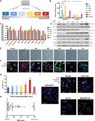
- Fig. 1. The heterogeneous cell line SUM149PT contains multiple distinct EMT states that can be isolated as single-cell clones. ( A ) A schematic of the flow cytometry method used to isolate single-cell clones that present as an epithelial (E), three distinct intermediate (EM1, EM2, and EM3), and two mesenchymal (M1 and M2) EMT states. ( B ) In vitro assessment of clonal migratory and invasive characteristics as measured in a standard transwell assay ( n = 3, SD, **** P < 0.0001, and * P < 0.05). Canonical EMT marker expression levels as determined by ( C ) quantitative RT-PCR (SD, n = 4) or ( D ) immunoblotting to rank SUM149 clones along the EMT spectrum. ( E ) Bright-field and ( F ) immunofluorescent images of EMT clones in vitro stained with vimentin and E-cadherin displaying cell morphology and marker expression and localization, respectively. ( G ) EMT signature of EMT clones and parental line generated from the ordinal multinomial logistic regression method of gene scoring and ( H ) distribution of EMT score of the EMT clones among other breast cancer cell lines from the CCLE. ( I ) Immunofluorescent staining for E-cadherin (red), vimentin (green), and KRT8 (white) of four triple-negative breast cancer lines (two intermediate, HCC38 and Cal-51; one epithelial, HCC1143; and one mesenchymal, MDA-MB-231) from the CCLE displaying heterogeneous phenotypes.