Explore
 Validate
 Learn
Learn Western blot
Antibody data
- Antibody Data
- Antigen structure
- References [1]
- Comments [0]
- Validations
- Western blot [5]
- Immunocytochemistry [1]
- Chromatin Immunoprecipitation [3]
- Other assay [2]
Submit
Validation data
Reference
Comment
Report error
- Product number
- PA5-31919 - Provider product page

- Provider
- Invitrogen Antibodies
- Product name
- SMYD3 Polyclonal Antibody
- Antibody type
- Polyclonal
- Antigen
- Recombinant protein fragment
- Description
- Recommended positive controls: DDDDK-tagged SMYD3-transfected 293T, 293T, A431, HeLa, HepG2, A549, SKOV3, HCT116.
- Concentration
- 1.23 mg/mL
Submitted references Small molecule inhibitors and CRISPR/Cas9 mutagenesis demonstrate that SMYD2 and SMYD3 activity are dispensable for autonomous cancer cell proliferation.
Thomenius MJ, Totman J, Harvey D, Mitchell LH, Riera TV, Cosmopoulos K, Grassian AR, Klaus C, Foley M, Admirand EA, Jahic H, Majer C, Wigle T, Jacques SL, Gureasko J, Brach D, Lingaraj T, West K, Smith S, Rioux N, Waters NJ, Tang C, Raimondi A, Munchhof M, Mills JE, Ribich S, Porter Scott M, Kuntz KW, Janzen WP, Moyer M, Smith JJ, Chesworth R, Copeland RA, Boriack-Sjodin PA
PloS one 2018;13(6):e0197372
PloS one 2018;13(6):e0197372
No comments: Submit comment
Supportive validation
- Submitted by
- Invitrogen Antibodies (provider)
- Main image

- Experimental details
- Western blot analysis of SMYD3 using A) 30 µg 293T whole cell lysate and B) 30 µg whole cell lysate of human SMYD3-transfected 293T cells (partial fragment). Samples were loaded onto a 10% SDS-PAGE gel and probed with a SMYD3 polyclonal antibody (Product # PA5-31919) at a dilution of 1:1000.

- Submitted by
- Invitrogen Antibodies (provider)
- Main image

- Experimental details
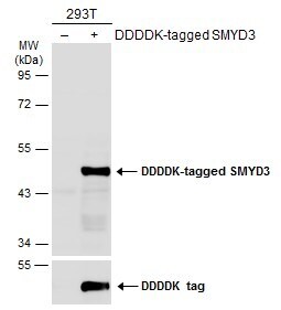
- Western Blot analysis of SMYD3 was performed by separating 30 µg of non-transfected (–) and transfected (+) 293T whole cell extracts by 10% SDS-PAGE. Proteins were transferred to a membrane and probed with a SMYD3 Polyclonal Antibody (Product # PA5-31919) at a dilution of 1:1000. The HRP-conjugated anti-rabbit IgG antibody was used to detect the primary antibody.
- Submitted by
- Invitrogen Antibodies (provider)
- Main image

- Experimental details
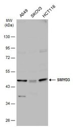
- SMYD3 Polyclonal Antibody detects SMYD3 protein by Western blot analysis. Various whole cell extracts (30 µg) were separated by 10% SDS-PAGE, and the membrane was blotted with SMYD3 Polyclonal Antibody (Product # PA5-31919) diluted at a dilution of 1:1,000.

- Submitted by
- Invitrogen Antibodies (provider)
- Main image
- Experimental details
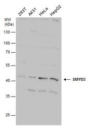
- Western Blot analysis of SMYD3 was performed by separating 30 µg of various whole cell extracts by 10% SDS-PAGE. Proteins were transferred to a membrane and probed with a SMYD3 Polyclonal Antibody (Product # PA5-31919) at a dilution of 1:1000 and a HRP-conjugated anti-rabbit IgG secondary antibody.

- Submitted by
- Invitrogen Antibodies (provider)
- Main image
- Experimental details
- Western Blot analysis of SMYD3 was performed by separating 30 µg of various whole cell extracts by 10% SDS-PAGE. Proteins were transferred to a membrane and probed with a SMYD3 Polyclonal Antibody (Product # PA5-31919) at a dilution of 1:1,000.

Supportive validation
- Submitted by
- Invitrogen Antibodies (provider)
- Main image

- Experimental details
- SMYD3 Polyclonal Antibody detects SMYD3 protein at cytoplasm by immunofluorescent analysis. Sample: HCT116 cells were fixed in ice-cold MeOH for 5 min. Green: SMYD3 protein stained by SMYD3 Polyclonal Antibody (Product # PA5-31919) diluted at 1:500. Blue: Hoechst 33342 staining.

Supportive validation
- Submitted by
- Invitrogen Antibodies (provider)
- Main image
- Experimental details
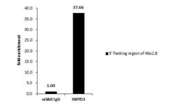
- Cross-linked ChIP was performed with HepG2 chromatin extract and 5 µg of either normal rabbit IgG or a SMYD3 polyclonal antibody (Product # PA5-31919). The precipitated DNA was detected by PCR with primer set targeting to 5' flanking region of Nkx2.8.

- Submitted by
- Invitrogen Antibodies (provider)
- Main image

- Experimental details
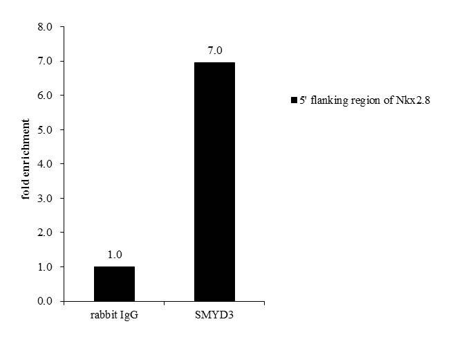
- Cross-linked ChIP analysis of SMYD3 in HepG2 chromatin extract using 5 µg of either normal rabbit IgG or anti-SMYD3 antibody (Product # PA5-31919). The precipitated DNA was detected by PCR with primer set targeting to 5' flanking region of Nkx2.8.

- Submitted by
- Invitrogen Antibodies (provider)
- Main image

- Experimental details
- Cross-linked ChIP was performed with HepG2 chromatin extract and 5 µg of either normal rabbit IgG or SMYD3 Polyclonal Antibody (Product # PA5-31919). The precipitated DNA was detected by PCR with primer set targeting to 5 flanking region of Nkx2.8.
Supportive validation
- Submitted by
- Invitrogen Antibodies (provider)
- Main image
- Experimental details
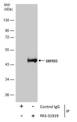
- Immunoprecipitation of SMYD3 was performed in 293T whole cell extracts using 5 µg of SMYD3 Polyclonal Antibody (Product # PA5-31919). Samples were transferred to a membrane and probed with SMYD3 Polyclonal Antibody as a primary antibody and an HRP-conjugated anti-Rabbit IgG was used as a secondary antibody.
- Submitted by
- Invitrogen Antibodies (provider)
- Main image
- Experimental details
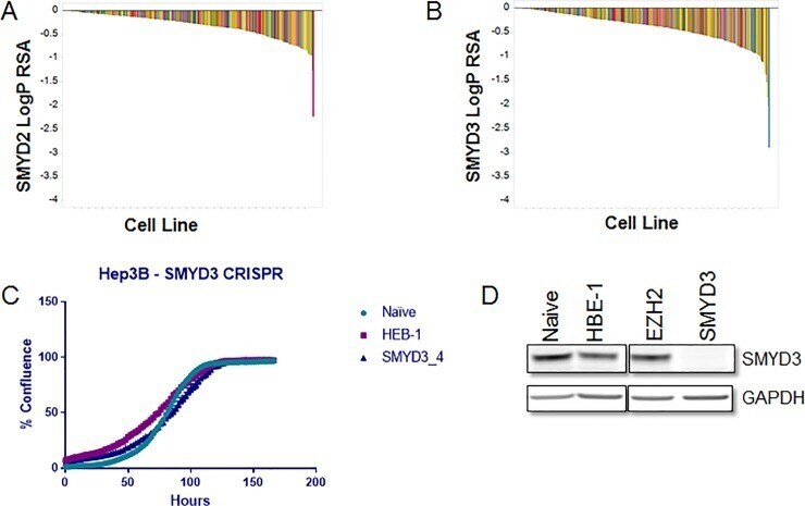
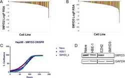
- Fig 5 Gene ablation techniques show no dependence on SMYD2 or SMYD3 for cancer cell proliferation. Waterfall plot representing LogP RSA scores for sgRNAs targeting A) SMYD2 and B) SMYD3. 313 cell lines were infected with a library of 6500 sgRNAs targeting 600 different genes. LogP RSA scores represent depletion of guides from an infected cell population. Each bar represents a different cell line. Bars are colored by cancer subtype. C) Percent confluency of Hep3B cells infected with CRISPR viruses containing CAS9 and sgRNAs targeting HBE-1, EZH2 (negative controls) or SMYD3. Cell density was evaluated using an Incucyte Zoom. Growth curves were initiated 24 days following virus infection and puromycin selection. Plotted data is the average of three biological replicates. Error bars represent standard deviation (not readily visible on scale). D) SMYD3 western blot of lysates derived from Hep3B cells infected with CAS9 and SMYD3 sgRNA. Parental Hep3Bs and Hep3Bs stably infected with HBE-1, EZH2 (negative controls) or SMYD3 were lysed and probed for SMYD3 levels by western. GAPDH levels were evaluated as a loading control.