Explore
 Validate
 Learn
Learn Western blot
Antibody data
- Antibody Data
- Antigen structure
- References [2]
- Comments [0]
- Validations
- Western blot [8]
- Immunoprecipitation [1]
- Immunohistochemistry [3]
Submit
Validation data
Reference
Comment
Report error
- Product number
- GTX118220 - Provider product page

- Provider
- GeneTex
- Proper citation
- GeneTex Cat#GTX118220, RRID:AB_10618954
- Product name
- SLIT2 antibody
- Antibody type
- Polyclonal
- Reactivity
- Human, Mouse, Rat
- Host
- Rabbit
Submitted references PlexinA1 is a new Slit receptor and mediates axon guidance function of Slit C-terminal fragments.
Down-regulation of Slit-Robo pathway mediating neuronal cytoskeletal remodeling processes facilitates the antidepressive-like activity of Gastrodia elata Blume.
Delloye-Bourgeois C, Jacquier A, Charoy C, Reynaud F, Nawabi H, Thoinet K, Kindbeiter K, Yoshida Y, Zagar Y, Kong Y, Jones YE, Falk J, Chédotal A, Castellani V
Nature neuroscience 2015 Jan;18(1):36-45
Nature neuroscience 2015 Jan;18(1):36-45
Down-regulation of Slit-Robo pathway mediating neuronal cytoskeletal remodeling processes facilitates the antidepressive-like activity of Gastrodia elata Blume.
Lin SH, Chen WC, Lu KH, Chen PJ, Hsieh SC, Pan TM, Chen ST, Sheen LY
Journal of agricultural and food chemistry 2014 Oct 29;62(43):10493-503
Journal of agricultural and food chemistry 2014 Oct 29;62(43):10493-503
No comments: Submit comment
Supportive validation
- Submitted by
- GeneTex (provider)
- Main image
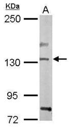
- Experimental details
- Sample (50 ?g of whole cell lysate) A: Mouse brain 5% SDS PAGE GTX118220 diluted at 1:1000 The HRP-conjugated anti-rabbit IgG antibody (GTX213110-01) was used to detect the primary antibody.

- Submitted by
- GeneTex (provider)
- Main image

- Experimental details
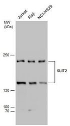
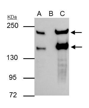
- SLIT2 antibody detects SLIT2 protein by western blot analysis.A. 30 ?g 293T whole cell lysate/extract B. 30 ?g A431 whole cell lysate/extract C. 30 ?g Jurkat whole cell lysate/extract D. 30 ?g Raji whole cell lysate/extract10% SDS-PAGESLIT2 antibody (GTX118220) dilution: 1:1000 The HRP-conjugated anti-rabbit IgG antibody (GTX213110-01) was used to detect the primary antibody.
- Submitted by
- GeneTex (provider)
- Main image
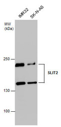
- Experimental details
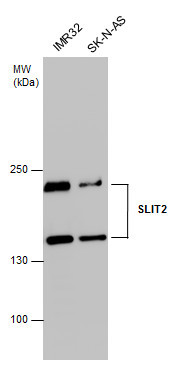
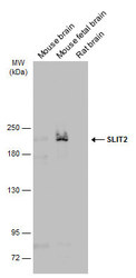
- SLIT2 antibody detects SLIT2 protein by western blot analysis. Various whole cell extracts (30 ?g) were separated by 5% SDS-PAGE, and the membrane was blotted with SLIT2 antibody (GTX118220) diluted at 1:1000. The HRP-conjugated anti-rabbit IgG antibody (GTX213110-01) was used to detect the primary antibody.
- Submitted by
- GeneTex (provider)
- Main image
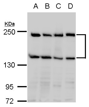
- Experimental details
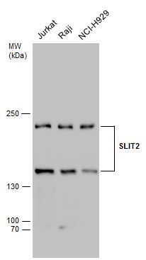
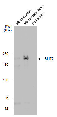
- Various whole cell extracts (30 ?g) were separated by 5% SDS-PAGE, and the membrane was blotted with SLIT2 antibody (GTX118220) diluted at 1:1000.
- Submitted by
- GeneTex (provider)
- Main image
- Experimental details
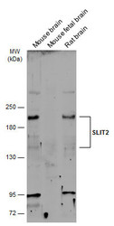
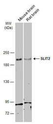
- Various tissue extracts (30 ?g) were separated by 5% SDS-PAGE, and the membrane was blotted with SLIT2 antibody (GTX118220) diluted at 1:500. The HRP-conjugated anti-rabbit IgG antibody (GTX213110-01) was used to detect the primary antibody.

- Submitted by
- GeneTex (provider)
- Main image
- Experimental details
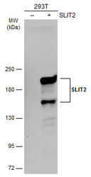
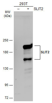
- Non-transfected (¡V) and transfected (+) 293T whole cell extracts (30 ?g) were separated by 5% SDS-PAGE, and the membrane was blotted with SLIT2 antibody (GTX118220) diluted at 1:4000. The HRP-conjugated anti-rabbit IgG antibody (GTX213110-01) was used to detect the primary antibody.
- Submitted by
- GeneTex (provider)
- Main image
- Experimental details
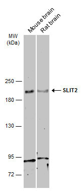
- Various tissue extracts (50 ?g) were separated by 5% SDS-PAGE, and the membrane was blotted with SLIT2 antibody (GTX118220) diluted at 1:500. The HRP-conjugated anti-rabbit IgG antibody (GTX213110-01) was used to detect the primary antibody.
- Submitted by
- GeneTex (provider)
- Main image
- Experimental details
- Various tissue extracts (30 ?g) were separated by 5% SDS-PAGE, and the membrane was blotted with SLIT2 antibody (GTX118220) diluted at 1:3000. The HRP-conjugated anti-rabbit IgG antibody (GTX213110-01) was used to detect the primary antibody.
Supportive validation
- Submitted by
- GeneTex (provider)
- Main image

- Experimental details
- SLIT2 antibody immunoprecipitates SLIT2 protein in IP experiments. IP Sample: Raji whole cell lysate/extract A : 30 £gg whole cell lysate/extract of SLIT2 protein expressing Raji cells B : Control with 3 £gg of pre-immune rabbit IgG C : Immunoprecipitation of SLIT2 by 3 £gg of SLIT2 antibody (GTX118220) 5% SDS-PAGE The immunoprecipitated SLIT2 protein was detected by SLIT2 antibody (GTX118220) diluted at 1 : 1000. EasyBlot anti-rabbit IgG (HRP) (GTX221666-01) was used as a secondary reagent.
Supportive validation
- Submitted by
- GeneTex (provider)
- Main image

- Experimental details
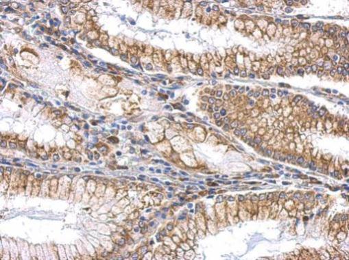
- SLIT2 antibody detects SLIT2 protein at cytosol and membrane on gastric carcinoma by immunohistochemical analysis. Sample: Paraffin-embedded human gastric carcinoma. SLIT2 antibody (GTX118220) dilution: 1:500.
- Submitted by
- GeneTex (provider)
- Main image
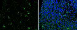
- Experimental details
- SLIT2 antibody detects SLIT2 protein at cytoplasm in mouse fetal brain by immunohistochemical analysis.Sample: Paraffin-embedded mouse fetal brain. Green: SLIT2 antibody (GTX118220) diluted at 1:200. The signal was developed using goat anti-rabbit IgG antibody (Dylight488) (GTX213110-04).Blue: Nuclear staining with Hoechst 33342.

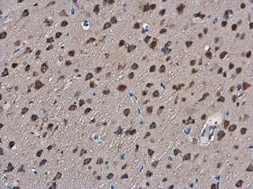
- Submitted by
- GeneTex (provider)
- Main image
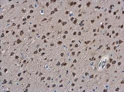
- Experimental details
- SLIT2 antibody detects SLIT2 protein at cytoplasm in rat brain by immunohistochemical analysis. Sample: Paraffin-embedded rat brain. SLIT2 antibody (GTX118220) diluted at 1:400.