 Explore
 Validate
 Learn
Learn Western blot
 Immunocytochemistry
Antibody data
- Antibody Data
- Antigen structure
- References [2]
- Comments [0]
- Validations
- Western blot [1]
- Immunocytochemistry [1]
- Immunohistochemistry [7]
Submit
Validation data
Reference
Comment
Report error
- Product number
- HPA000248 - Provider product page

- Provider
- Atlas Antibodies
- Proper citation
- Atlas Antibodies Cat#HPA000248, RRID:AB_1079507
- Product name
- Anti-NSDHL
- Antibody type
- Polyclonal
- Reactivity
- Human
- Host
- Rabbit
- Conjugate
- Unconjugated
- Antigen sequence
MEPAVSEPMRDQVARTHLTEDTPKVNADIEKVNQN
QAKRCTVIGGSGFLGQHMVEQLLARGYAVNVFDIQ
QGFDNPQVRFFLGDLCSRQDLYPALKGVNTVFHCA
SPPPSSNNKELFYRVNYIGT- Isotype
- IgG
- Vial size
- 100 µl
- Storage
- Store at +4°C for short term storage. Long time storage is recommended at -20°C.
Submitted references Immunofluorescence and fluorescent-protein tagging show high correlation for protein localization in mammalian cells.
Proteomic analysis of enriched lysosomes at early phase of camptothecin-induced apoptosis in human U-937 cells.
Stadler C, Rexhepaj E, Singan VR, Murphy RF, Pepperkok R, Uhlén M, Simpson JC, Lundberg E
Nature methods 2013 Apr;10(4):315-23
Nature methods 2013 Apr;10(4):315-23
Proteomic analysis of enriched lysosomes at early phase of camptothecin-induced apoptosis in human U-937 cells.
Parent N, Winstall E, Beauchemin M, Paquet C, Poirier GG, Bertrand R
Journal of proteomics 2009 Aug 20;72(6):960-73
Journal of proteomics 2009 Aug 20;72(6):960-73
No comments: Submit comment
Enhanced validation
- Submitted by
- Atlas Antibodies (provider)
- Enhanced method
- Independent antibody validation
- Main image
- Experimental details
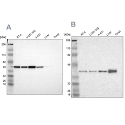
- Western blot analysis using Anti-NSDHL antibody HPA000248 (A) shows similar pattern to independent antibody HPA000571 (B).

Supportive validation
- Submitted by
- Atlas Antibodies (provider)
- Main image

- Experimental details
- Immunofluorescent staining of human cell line A-431 shows localization to endoplasmic reticulum & lipid droplets.
- Sample type
- HUMAN

Enhanced validation
Enhanced validation
Supportive validation
- Submitted by
- Atlas Antibodies (provider)
- Enhanced method
- Orthogonal validation
- Main image

- Experimental details
- Immunohistochemistry analysis in human epididymis and lymph node tissues using Anti-NSDHL antibody. Corresponding NSDHL RNA-seq data are presented for the same tissues.
- Sample type
- HUMAN

Enhanced validation
- Submitted by
- Atlas Antibodies (provider)
- Enhanced method
- Independent antibody validation
- Main image

- Experimental details
- Immunohistochemical staining of human cerebral cortex, epididymis, lymph node and testis using Anti-NSDHL antibody HPA000248 (A) shows similar protein distribution across tissues to independent antibody HPA000571 (B).

Supportive validation
- Submitted by
- Atlas Antibodies (provider)
- Main image

- Experimental details
- Immunohistochemical staining of human testis shows cytoplasmic positivity in Leydig cells.

- Submitted by
- Atlas Antibodies (provider)
- Main image

- Experimental details
- Immunohistochemical staining of human epididymis shows high expression.
- Sample type
- HUMAN

- Submitted by
- Atlas Antibodies (provider)
- Main image

- Experimental details
- Immunohistochemical staining of human lymph node shows low expression as expected.
- Sample type
- HUMAN

- Submitted by
- Atlas Antibodies (provider)
- Main image

- Experimental details
- Immunohistochemical staining of human testis using Anti-NSDHL antibody HPA000248.
- Sample type
- HUMAN

- Submitted by
- Atlas Antibodies (provider)
- Main image

- Experimental details
- Immunohistochemical staining of human cerebral cortex using Anti-NSDHL antibody HPA000248.
- Sample type
- HUMAN
