Explore
 Validate
 Learn
Learn Western blot
 ELISA
ELISA Immunocytochemistry
Antibody data
- Antibody Data
- Antigen structure
- References [0]
- Comments [0]
- Validations
- Immunocytochemistry [1]
- Flow cytometry [1]
Submit
Validation data
Reference
Comment
Report error
- Product number
- MA5-14674 - Provider product page

- Provider
- Invitrogen Antibodies
- Product name
- Anti-CEA Monoclonal Antibody (1105)
- Antibody type
- Monoclonal
- Antigen
- Purifed from natural sources
- Description
- The predicted MW of CEA is ~77kD, but by Western blot MA5-14675 detects CEA with varying degrees of glycosylation at ~77-180kD. Product MA514675 is a smaller package size of MIC0102 (formerly sold as a Seradyn product).
- Reactivity
- Human
- Host
- Mouse
- Isotype
- IgG
- Antibody clone number
- 1105
- Vial size
- 100 ug
- Concentration
- 1 mg/ml
- Storage
- -20° C, Avoid Freeze/Thaw Cycles
No comments: Submit comment
Supportive validation
- Submitted by
- Invitrogen Antibodies (provider)
- Main image
- Experimental details
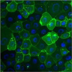
- Immunofluorescent analysis of Carcinoembryonic Antigen (CEA, green) in BxPC-3 cells. Cells were fixed with 4% paraformaldehyde, permeabilized with 0.1% Triton X-100, and blocked with 0.3% BSA in PBS, each for 15 minutes at room temperature. Cells were stained with a CEA monoclonal antibody (Product # MA5-14674) at a dilution of 10 µg/mL in blocking buffer for 1 hour at room temperature, and then incubated with a goat anti-mouse IgG Superclonal™ secondary antibody, Alexa Fluor® 488 conjugate (Product # A28175) at a dilution of 1:1000 for 1 hour at room temperature. Nuclei (blue) were stained with Hoechst nuclear stain. Images were taken on a Thermo Scientific ToxInsight Instrument at 20X magnification.

Supportive validation
- Submitted by
- Invitrogen Antibodies (provider)
- Main image

- Experimental details
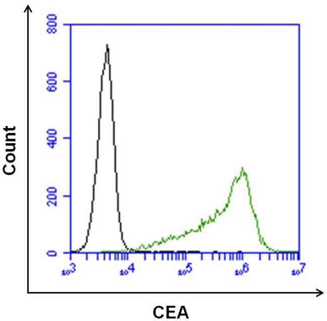
- Flow cytometry analysis of Carcinoembryonic Antigen (CEA) on BxPC-3 cells. Cells were harvested with 0.25% Trypsin-EDTA and stained with a CEA monoclonal antibody (Product # MA5-14674) at a dilution of 10 µg/mL (pink histogram), or with a mouse isotype control (black histogram) at a dilution of 10 µg/mL in 5% FCS in PBS. After incubation of the primary antibody for 1 hour on ice, the cells were stained with a goat anti-mouse IgG secondary antibody, DyLight 488 conjugate (Product # 35502) at a dilution of 1:40 for 1 hour on ice. A representative 10,000 cells were acquired for each sample.