 Explore
 Validate
 Learn
Learn Western blot
 Immunocytochemistry
Antibody data
- Antibody Data
- Antigen structure
- References [9]
- Comments [0]
- Validations
- Western blot [6]
- Immunohistochemistry [2]
- Flow cytometry [2]
Submit
Validation data
Reference
Comment
Report error
- Product number
- NBP2-29415 - Provider product page

- Provider
- Novus Biologicals
- Proper citation
- Novus Cat#NBP2-29415, RRID:AB_2631231
- Product name
- Mouse Monoclonal GFAP Antibody
- Antibody type
- Monoclonal
- Description
- Protein A or G purified. This MAb recognizes a protein of ~50kDa which is identified as Glial Fibrillary Acidic Protein (GFAP). It shows no cross-reaction with other intermediate filament proteins. GFAP is specifically found in astroglia. GFAP is a very popular marker for localizing benign astrocyte and neoplastic cells of glial origin in the central nervous system. Antibody to GFAP is useful in differentiating primary gliomas from metastatic lesions in the brain and for documenting astrocytic differentiation in tumors outside the CNS.
- Reactivity
- Human, Mouse, Rat, Bovine, Chicken/Avian, Porcine, Rabbit
- Host
- Mouse
- Isotype
- IgG
- Vial size
- 0.1 mg
- Concentration
- 0.2 mg/ml
- Storage
- Store at 4C.
Submitted references Evaluation of the NAD(+) biosynthetic pathway in ALS patients and effect of modulating NAD(+) levels in hSOD1-linked ALS mouse models.
A Method to Visualize the Nanoscopic Morphology of Astrocytes In Vitro and In Situ.
Transfer and Integration of Breast Milk Stem Cells to the Brain of Suckling Pups.
Folate homeostasis in epileptic rats.
Probing nano-organization of astroglia with multi-color super-resolution microscopy.
Nanoparticle fullerol alleviates radiculopathy via NLRP3 inflammasome and neuropeptides.
Tracking inflammation in the epileptic rat brain by bi-functional fluorescent and magnetic nanoparticles.
Morphological plasticity of astroglia: Understanding synaptic microenvironment.
The effects of apelin on the electrical activity of hypothalamic magnocellular vasopressin and oxytocin neurons and somatodendritic Peptide release.
Harlan BA, Killoy KM, Pehar M, Liu L, Auwerx J, Vargas MR
Experimental neurology 2020 May;327:113219
Experimental neurology 2020 May;327:113219
A Method to Visualize the Nanoscopic Morphology of Astrocytes In Vitro and In Situ.
Heller JP, Rusakov DA
Methods in molecular biology (Clifton, N.J.) 2019;1938:69-84
Methods in molecular biology (Clifton, N.J.) 2019;1938:69-84
Transfer and Integration of Breast Milk Stem Cells to the Brain of Suckling Pups.
Aydın MŞ, Yiğit EN, Vatandaşlar E, Erdoğan E, Öztürk G
Scientific reports 2018 Sep 24;8(1):14289
Scientific reports 2018 Sep 24;8(1):14289
Folate homeostasis in epileptic rats.
Mann A, Portnoy E, Han H, Inbar D, Blatch D, Shmuel M, Ben-Hur T, Eyal S, Ekstein D
Epilepsy research 2018 May;142:64-72
Epilepsy research 2018 May;142:64-72
Probing nano-organization of astroglia with multi-color super-resolution microscopy.
Heller JP, Michaluk P, Sugao K, Rusakov DA
Journal of neuroscience research 2017 Nov;95(11):2159-2171
Journal of neuroscience research 2017 Nov;95(11):2159-2171
Nanoparticle fullerol alleviates radiculopathy via NLRP3 inflammasome and neuropeptides.
Jin L, Ding M, Oklopcic A, Aghdasi B, Xiao L, Li Z, Jevtovic-Todorovic V, Li X
Nanomedicine : nanotechnology, biology, and medicine 2017 Aug;13(6):2049-2059
Nanomedicine : nanotechnology, biology, and medicine 2017 Aug;13(6):2049-2059
Tracking inflammation in the epileptic rat brain by bi-functional fluorescent and magnetic nanoparticles.
Portnoy E, Polyak B, Inbar D, Kenan G, Rai A, Wehrli SL, Roberts TP, Bishara A, Mann A, Shmuel M, Rozovsky K, Itzhak G, Ben-Hur T, Magdassi S, Ekstein D, Eyal S
Nanomedicine : nanotechnology, biology, and medicine 2016 Jul;12(5):1335-45
Nanomedicine : nanotechnology, biology, and medicine 2016 Jul;12(5):1335-45
Morphological plasticity of astroglia: Understanding synaptic microenvironment.
Heller JP, Rusakov DA
Glia 2015 Dec;63(12):2133-51
Glia 2015 Dec;63(12):2133-51
The effects of apelin on the electrical activity of hypothalamic magnocellular vasopressin and oxytocin neurons and somatodendritic Peptide release.
Tobin VA, Bull PM, Arunachalam S, O'Carroll AM, Ueta Y, Ludwig M
Endocrinology 2008 Dec;149(12):6136-45
Endocrinology 2008 Dec;149(12):6136-45
No comments: Submit comment
Supportive validation
- Submitted by
- Novus Biologicals (provider)
- Main image
- Experimental details
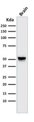
- Western Blot: GFAP Antibody (GA5) [NBP2-29415] - Analysis of GFAP in human brain lysate using GFAP (GA5) antibody at 1 ug/ml. goat anti-mouse Ig HRP secondary antibody and PicoTect ECL substrate solution were used for this test.

- Submitted by
- Novus Biologicals (provider)
- Main image

- Experimental details
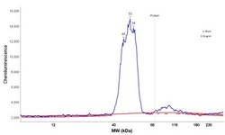
- Simple Western: GFAP Antibody (GA5) [NBP2-29415] - Simple Western lane view shows a specific band for GFAP in 0.2 mg/ml of h. Brain lysate(s). This experiment was performed under reducing conditions using the 12-230 kDa separation system.

- Submitted by
- Novus Biologicals (provider)
- Main image

- Experimental details
- Simple Western: GFAP Antibody (GA-5) [NBP2-29415] - Simple Western lane view shows a specific band for GFAP in 0.2 mg/ml of h. Brain lysate(s). This experiment was performed under reducing conditions using the 12-230 kDa separation system.

- Submitted by
- Novus Biologicals (provider)
- Main image
- Experimental details
- Simple Western: GFAP Antibody (GA-5) [NBP2-29415] - Electropherogram image of the corresponding Simple Western lane. GFAP antibody was used at 10 ug/ml dilution of h. Brain lysates(s) respectively.

- Submitted by
- Novus Biologicals (provider)
- Main image
- Experimental details
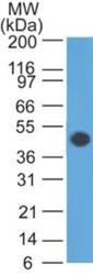
- Western Blot: GFAP Antibody (GA-5) [NBP2-29415] - Western Blot Analysis of human brain tissue lysate using GFAP Antibody (GA-5).

- Submitted by
- Novus Biologicals (provider)
- Main image

- Experimental details
- Western Blot: GFAP Antibody (GA-5) [NBP2-29415] - Analysis of GFAP in human brain lysate using GFAP (GA5) antibody at 1 ug/ml. goat anti-mouse Ig HRP secondary antibody and PicoTect ECL substrate solution were used for this test.

Supportive validation
- Submitted by
- Novus Biologicals (provider)
- Main image
- Experimental details
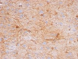
- Immunohistochemistry-Paraffin: GFAP Antibody (GA-5) [NBP2-29415] - Formalin-paraffin human brain stained with GFAP Ab (GA-5). Note cytoplasmic staining.

- Submitted by
- Novus Biologicals (provider)
- Main image

- Experimental details
- Immunohistochemistry-Paraffin: GFAP Antibody (GA-5) [NBP2-29415] - Formalin-fixed, paraffin-embedded human Cerebellum stained with GFAP Antibody (GA-5).

Supportive validation
- Submitted by
- Novus Biologicals (provider)
- Main image

- Experimental details
- Flow Cytometry: GFAP Antibody (GA-5) [NBP2-29415] - Experimental autoimmune encephalomyelitis was induced in C57BL6/J mice, and mononuclear cells were isolated from the CNS at day 10 (onset of symptoms). Cells were stained for GFAP, Neun, CX3CL1, CXCL12, CCL2, CD45 and CD11b, plus for viability to exclude dead cells. GFAP staining is shown for viable cells. Image from verified customer review.

- Submitted by
- Novus Biologicals (provider)
- Main image

- Experimental details
- Flow Cytometry: GFAP Antibody (GA-5) [NBP2-29415] - Flow Cytometric Analysis of T98G cells using GFAP Antibody (GA-5) followed by Goat anti-Mouse IgG-CF488 (Blue); Isotype Control (Red).
