 Explore
 Validate
 Learn
Learn Western blot
 Immunohistochemistry
Antibody data
- Antibody Data
- Antigen structure
- References [10]
- Comments [0]
- Validations
- Immunohistochemistry [3]
- Other assay [5]
Submit
Validation data
Reference
Comment
Report error
- Product number
- OPA1-06100 - Provider product page

- Provider
- Invitrogen Antibodies
- Product name
- GFAP Polyclonal Antibody
- Antibody type
- Polyclonal
- Antigen
- Other
- Description
- OPA1-06100 detects glial fibrillary acid protein (GFAP) from human, mouse, zebrafish, and rat tissues. OPA1-06100 has been successfully used in Western blot and immunohistochemistry (paraffin and frozen) procedures. By Western blot, this antibody detects a single protein representing GFAP rat brain homogenate. Immunohistochemical staining of GFAP in rat brain with OPA1-6100 results in intense staining of astrocytes and Schwann cells. The OPA1-06100 immunogen is cytoskeletal preparation of human spinal cord. Store at 4ºC or in small aliquots at -20ºC.
- Reactivity
- Human, Mouse, Rat, Zebrafish
- Host
- Rabbit
- Isotype
- IgG
- Vial size
- 100 µL
- Concentration
- Conc. Not Determined
- Storage
- Store at 4°C short term. For long term storage, store at -20°C, avoiding freeze/thaw cycles.
Submitted references Insulin Diminishes Superoxide Increase in Cytosol and Mitochondria of Cultured Cortical Neurons Treated with Toxic Glutamate.
The TGFβ/Notch axis facilitates Müller cell-to-epithelial transition to ultimately form a chronic glial scar.
Full-length Dhh and N-terminal Shh act as competitive antagonists to regulate angiogenesis and vascular permeability.
Lasting alterations induced in glial cell phenotypes by short exposure to alcohol during embryonic development in zebrafish.
Desert Hedgehog-Driven Endothelium Integrity Is Enhanced by Gas1 (Growth Arrest-Specific 1) but Negatively Regulated by Cdon (Cell Adhesion Molecule-Related/Downregulated by Oncogenes).
Targeting Brain Disease in MPSII: Preclinical Evaluation of IDS-Loaded PLGA Nanoparticles.
A novel fluorescent imaging technique for assessment of cerebral vasospasm after experimental subarachnoid hemorrhage.
The nucleotide-binding leucine-rich repeat (NLR) family member NLRX1 mediates protection against experimental autoimmune encephalomyelitis and represses macrophage/microglia-induced inflammation.
Miz1 is required to maintain autophagic flux.
Behavioral and neurochemical alterations in mice deficient in anaplastic lymphoma kinase suggest therapeutic potential for psychiatric indications.
Pinelis V, Krasilnikova I, Bakaeva Z, Surin A, Boyarkin D, Fisenko A, Krasilnikova O, Pomytkin I
International journal of molecular sciences 2022 Oct 20;23(20)
International journal of molecular sciences 2022 Oct 20;23(20)
The TGFβ/Notch axis facilitates Müller cell-to-epithelial transition to ultimately form a chronic glial scar.
Conedera FM, Pousa AMQ, Mercader N, Tschopp M, Enzmann V
Molecular neurodegeneration 2021 Sep 30;16(1):69
Molecular neurodegeneration 2021 Sep 30;16(1):69
Full-length Dhh and N-terminal Shh act as competitive antagonists to regulate angiogenesis and vascular permeability.
Hollier PL, Chapouly C, Diop A, Guimbal S, Cornuault L, Gadeau AP, Renault MA
Cardiovascular research 2021 Nov 1;117(12):2489-2501
Cardiovascular research 2021 Nov 1;117(12):2489-2501
Lasting alterations induced in glial cell phenotypes by short exposure to alcohol during embryonic development in zebrafish.
Chatterjee D, Mahabir S, Chatterjee D, Gerlai R
Addiction biology 2021 Jan;26(1):e12867
Addiction biology 2021 Jan;26(1):e12867
Desert Hedgehog-Driven Endothelium Integrity Is Enhanced by Gas1 (Growth Arrest-Specific 1) but Negatively Regulated by Cdon (Cell Adhesion Molecule-Related/Downregulated by Oncogenes).
Chapouly C, Hollier PL, Guimbal S, Cornuault L, Gadeau AP, Renault MA
Arteriosclerosis, thrombosis, and vascular biology 2020 Dec;40(12):e336-e349
Arteriosclerosis, thrombosis, and vascular biology 2020 Dec;40(12):e336-e349
Targeting Brain Disease in MPSII: Preclinical Evaluation of IDS-Loaded PLGA Nanoparticles.
Rigon L, Salvalaio M, Pederzoli F, Legnini E, Duskey JT, D'Avanzo F, De Filippis C, Ruozi B, Marin O, Vandelli MA, Ottonelli I, Scarpa M, Tosi G, Tomanin R
International journal of molecular sciences 2019 Apr 24;20(8)
International journal of molecular sciences 2019 Apr 24;20(8)
A novel fluorescent imaging technique for assessment of cerebral vasospasm after experimental subarachnoid hemorrhage.
Aum DJ, Vellimana AK, Singh I, Milner E, Nelson JW, Han BH, Zipfel GJ
Scientific reports 2017 Aug 22;7(1):9126
Scientific reports 2017 Aug 22;7(1):9126
The nucleotide-binding leucine-rich repeat (NLR) family member NLRX1 mediates protection against experimental autoimmune encephalomyelitis and represses macrophage/microglia-induced inflammation.
Eitas TK, Chou WC, Wen H, Gris D, Robbins GR, Brickey J, Oyama Y, Ting JP
The Journal of biological chemistry 2014 Feb 14;289(7):4173-9
The Journal of biological chemistry 2014 Feb 14;289(7):4173-9
Miz1 is required to maintain autophagic flux.
Wolf E, Gebhardt A, Kawauchi D, Walz S, von Eyss B, Wagner N, Renninger C, Krohne G, Asan E, Roussel MF, Eilers M
Nature communications 2013;4:2535
Nature communications 2013;4:2535
Behavioral and neurochemical alterations in mice deficient in anaplastic lymphoma kinase suggest therapeutic potential for psychiatric indications.
Bilsland JG, Wheeldon A, Mead A, Znamenskiy P, Almond S, Waters KA, Thakur M, Beaumont V, Bonnert TP, Heavens R, Whiting P, McAllister G, Munoz-Sanjuan I
Neuropsychopharmacology : official publication of the American College of Neuropsychopharmacology 2008 Feb;33(3):685-700
Neuropsychopharmacology : official publication of the American College of Neuropsychopharmacology 2008 Feb;33(3):685-700
No comments: Submit comment
Supportive validation
- Submitted by
- Invitrogen Antibodies (provider)
- Main image
- Experimental details
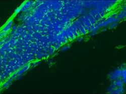
- Immunofluorescent analysis of 9 days old zebrafish embryo using GFAP polyclonal antibody (Product # OPA1-06100).

- Submitted by
- Invitrogen Antibodies (provider)
- Main image

- Experimental details
- Immunofluorescent analysis of 9 days old zebrafish embryo using GFAP polyclonal antibody (Product # OPA1-06100).

- Submitted by
- Invitrogen Antibodies (provider)
- Main image
- Experimental details
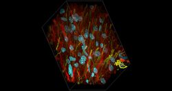
- Deep tissue imaging with non-curing, refractive index matched SlowFade™ Glass. Cryo-preserved rat brain sections (100 µm thick), stained for tubulin (red) with Mouse Anti-Beta3-Tubulin (Cat. No. MA1-118) and GFAP (yellow) Rabbit Anti-GFAP (Cat. No. OPA1-06100). Targets were detected with Alexa Fluor™ Plus 594 Goat Anti-Mouse (Cat. No. A-11032) and Alexa Fluor™ Plus 647 Goat Anti-Rabbit (Cat. No. A-32733). Nuclei (Cyan) were stained with DAPI (Cat. No. D1306). Slides were mounted with SlowFade™ Glass non-curing Antifade Mountant (Cat. No. S36917) and imaged with a Zeiss LSM 710 confocal microscope using a Plan-Apochromat 63×/1.4 NA Oil immersion objective at a rate of 71 nm in the x and y dimensions and 110 nm in the z dimension, with a pixel size of 0.07 µm. Z-projections were generated using Zeiss™ Zen software.

Supportive validation
- Submitted by
- Invitrogen Antibodies (provider)
- Main image
- Experimental details
- NULL
- Submitted by
- Invitrogen Antibodies (provider)
- Main image

- Experimental details
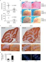
- Figure 6 Detection of neuroinflammation after SAH. Sections of ROX SE-perfused brains from mice that underwent sham and SAH surgeries were subjected to fluorescent immunohistochemical staining using anti-alpha-glial acidic fibrillary protein (GFAP) and anti-CD45 antibodies. A marked increase in astrocyte ( A ) and microglial activation ( B ) is observed after SAH. Scale bar: 50 mum.

- Submitted by
- Invitrogen Antibodies (provider)
- Main image

- Experimental details
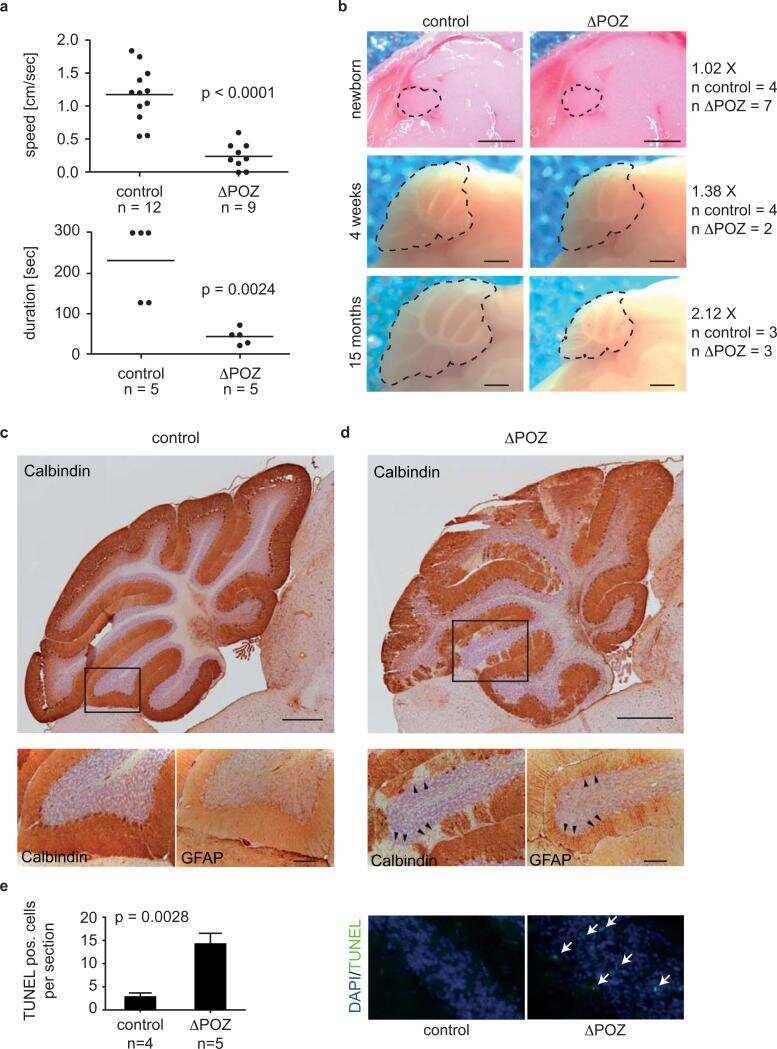
- Fig. 5 Human MCs show epithelial phenotype associated with TGFbeta/Notch under pathological condition. (A) Human H&E stained sections of retinas that show healthy cuboidal RPE (Neg) and retinas presenting drusen beneath the pigment epithelium (Drusen pos). Zoom-in view showing a healthy cuboidal RPE layer (Neg, top left corner) and drusen or micro drusen underneath the RPE layer (Drusen pos, top left corner). (B) Quantification of drusen as either hyalinized rounded deposits (> 25 mum) or micro drusen occurring singly or in a row (< 25 mum) in between the RPE and the Bruch's membrane. The analyzed length of the retina was 950 mum retina sections corresponding to the field of view. (C) Table summarizing the eight selected samples (total analyzed sections = 52) selected for H&E and immunofluorescence analysis. (D-J) Analysis of MC reactivity and phenotype of healthy retinas (Neg) and retinas presenting drusen (Drusen pos). Detection of GFAP (D.i-D.iv, E.i-E.iv), PCNA (F.i-F.iv, G.i-G.iv) and E-cadherin (H.i-H.iv, I.i-I.iv) in GS+ MCs. Shown are representative sections stained for GS and SOX9 (red), GFAP, PCNA and E-Cadherin (green). (J) Histogram illustrating the mean +- SD of the number of GFAP+, PCNA+ and E-cadherin+ cells normalized by the total number of GS+ cells expressed in percentage. Significant differences (****p < 0.0001) between ""Neg"" and ""Drusen pos"" retinas were determined by using a post-hoc Bonferroni two-way ANOVA test ( n = 8). (K-O) Analysis of regulators

- Submitted by
- Invitrogen Antibodies (provider)
- Main image

- Experimental details
- 6 FIGURE Zebrafish hindbrain stained with anti-GFAP antibody. Left most panel shows the schematic diagrams of the corresponding brain areas from where photomicrographs were taken. First column of photomicrographs shows control adult zebrafish hind brain; second column--DAPI stain applied to the corresponding area; and third column--superimposition of anti-GFAP antibody and DAPI stained photomicrographs; fourth column--adult zebrafish that were exposed to 1% alcohol during embryonic development--hindbrain, stained with anti-GFAP antibody; fifth column--DAPI stain applied to the corresponding area; sixth column--superimposition of anti-GFAP antibody and DAPI stained photomicrographs. The embryonic alcohol concentration (0 or 1% vol/vol) employed is indicated above the columns of photomicrographs. Magnification of the photomicrographs is also indicated

- Submitted by
- Invitrogen Antibodies (provider)
- Main image

- Experimental details
- 1 FIGURE Validation of the anti-GFAP (A) and anti-MBP (B) antibodies, employed in this study, by western blot analysis. Panel C shows the specificity of anti Galactocerebroside antibody used in this study by TLC immune-overlay blot technique. Source of samples is indicated above the corresponding lanes
