Explore
 Validate
 Learn
Learn Western blot
Antibody data
- Antibody Data
- Antigen structure
- References [4]
- Comments [0]
- Validations
- Western blot [3]
- ELISA [1]
- Other assay [2]
Submit
Validation data
Reference
Comment
Report error
- Product number
- PA5-18598 - Provider product page

- Provider
- Invitrogen Antibodies
- Product name
- GFAP Polyclonal Antibody
- Antibody type
- Polyclonal
- Antigen
- Synthetic peptide
- Description
- This antibody is predicted to react with canine and rat based on sequence homology.
- Reactivity
- Human, Mouse, Rat
- Host
- Goat
- Isotype
- IgG
- Vial size
- 100 µg
- Concentration
- 0.5 mg/mL
- Storage
- -20° C, Avoid Freeze/Thaw Cycles
Submitted references Metabolic Profiling of Suprachiasmatic Nucleus Reveals Multifaceted Effects in an Alzheimer's Disease Mouse Model.
Beneficial contribution of induced pluripotent stem cell-progeny to Connexin 47 dynamics during demyelination-remyelination.
Three-dimensional brain-on-chip model using human iPSC-derived GABAergic neurons and astrocytes: Butyrylcholinesterase post-treatment for acute malathion exposure.
Zika Virus Targets Glioblastoma Stem Cells through a SOX2-Integrin α(v)β(5) Axis.
Eeza MNH, Singer R, Höfling C, Matysik J, de Groot HJM, Roβner S, Alia A
Journal of Alzheimer's disease : JAD 2021;81(2):797-808
Journal of Alzheimer's disease : JAD 2021;81(2):797-808
Beneficial contribution of induced pluripotent stem cell-progeny to Connexin 47 dynamics during demyelination-remyelination.
Mozafari S, Deboux C, Laterza C, Ehrlich M, Kuhlmann T, Martino G, Baron-Van Evercooren A
Glia 2021 May;69(5):1094-1109
Glia 2021 May;69(5):1094-1109
Three-dimensional brain-on-chip model using human iPSC-derived GABAergic neurons and astrocytes: Butyrylcholinesterase post-treatment for acute malathion exposure.
Liu L, Koo Y, Russell T, Gay E, Li Y, Yun Y
PloS one 2020;15(3):e0230335
PloS one 2020;15(3):e0230335
Zika Virus Targets Glioblastoma Stem Cells through a SOX2-Integrin α(v)β(5) Axis.
Zhu Z, Mesci P, Bernatchez JA, Gimple RC, Wang X, Schafer ST, Wettersten HI, Beck S, Clark AE, Wu Q, Prager BC, Kim LJY, Dhanwani R, Sharma S, Garancher A, Weis SM, Mack SC, Negraes PD, Trujillo CA, Penalva LO, Feng J, Lan Z, Zhang R, Wessel AW, Dhawan S, Diamond MS, Chen CC, Wechsler-Reya RJ, Gage FH, Hu H, Siqueira-Neto JL, Muotri AR, Cheresh DA, Rich JN
Cell stem cell 2020 Feb 6;26(2):187-204.e10
Cell stem cell 2020 Feb 6;26(2):187-204.e10
No comments: Submit comment
Supportive validation
- Submitted by
- Invitrogen Antibodies (provider)
- Main image
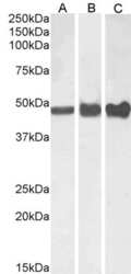
- Experimental details
- Western blot analysis of GFAP by a GFAP monoclonal antibody (Product # PA5-18598) at a concentration of 0.01 µg/mL. Human (A), Mouse (B) and (0.003 µg/mL) Rat (C) Brain lysate (35µg protein in RIPA buffer). Detected by chemiluminescence.
- Submitted by
- Invitrogen Antibodies (provider)
- Main image

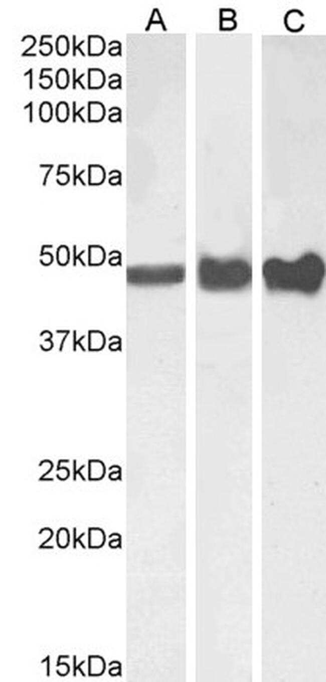
- Experimental details
- Western Blot staining of Mouse Brain lysate using Product # PA5-18598 at a concentration of 0.1 µg/mL, the primary antibody incubation was 1 hour and the detection method was chemiluminescence.

- Submitted by
- Invitrogen Antibodies (provider)
- Main image

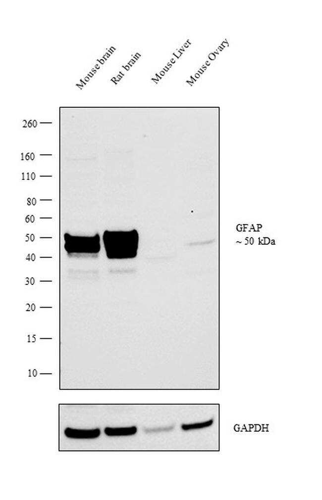
- Experimental details
- Western blot analysis was performed on tissue extracts (30 µg lysate) of Mouse brain (Lane 1), Rat brain (Lane 2), Mouse Liver (Lane 3) and Mouse Ovary (Lane 4). The blot was probed with Anti-GFAP Polyclonal Antibody (Product # PA5-18598, 1:1000 dilution) and detected by chemiluminescence using Rabbit anti-Goat IgG (H+L) Superclonal™ Secondary Antibody, HRP conjugate (Product # A27014, 0.25 µg/mL, 1:4000 dilution). A 50 kDa band corresponding to GFAP was observed in the Mouse and Rat brain, while it was not detected in Mouse Liver and Mouse Ovary.
Supportive validation
- Submitted by
- Invitrogen Antibodies (provider)
- Main image
- Experimental details
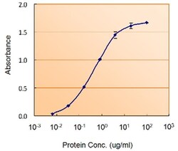
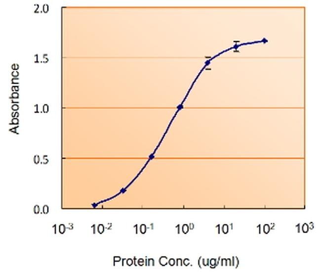
- ELIZA analysis of GFAP concentration using a GFAP antibody (Product # PA5-18598) (1.5µg/mL) as the reporter with a capture rabbit antibody (5µg/mL).
Supportive validation
- Submitted by
- Invitrogen Antibodies (provider)
- Main image
- Experimental details
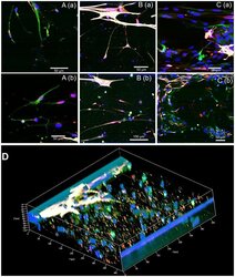
- 335.g003 Fig 3 Functional synapses between cells. A. Synapses between neurons (A1/N4); B. Synapses between neurons and astrocytes (A1/N4); C. No synapses in A4/N1 group. D. 3D co-culture with synapses (A1/N4). Red: Synaptophysin for synapses, green: beta-tubulin III for neurons, white: GFAP for astrocytes, and blue: Hoechst for nuclei.

- Submitted by
- Invitrogen Antibodies (provider)
- Main image
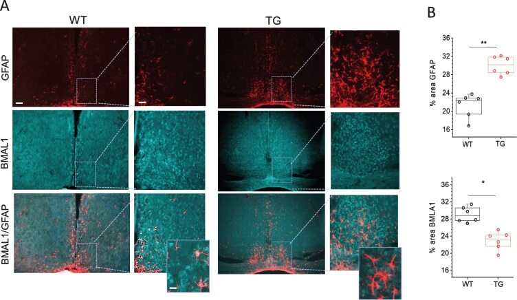
- Experimental details
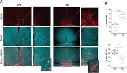
- Fig. 3 Immunohistochemical analyses of Bmal1 and GFAP staining in SCN of Tg2576 (TG) and wild-type (WT) mice. A) Representative confocal images of Bmal1 and GFAP stained sections through SCN of 18-month-old WT and TG mice. Scale bar, 250mum (first and third column); 60mum (second and fourth column) and 20mum (in magnifications). B) Quantitative analysis of Bmal1 and GFAP staining in SCN of 18 months old WT and Tg2576 (TG) mice ( n = 6 per group). ** p < 0.01, * p < 0.05. GFAP, glial fibrillary acidic protein.