 Explore
 Validate
 Learn
Learn Immunocytochemistry
 Immunohistochemistry
Antibody data
- Antibody Data
- Antigen structure
- References [2]
- Comments [0]
- Validations
- Immunocytochemistry [1]
Submit
Validation data
Reference
Comment
Report error
- Product number
- MA1-35377 - Provider product page

- Provider
- Invitrogen Antibodies
- Product name
- GFAP Monoclonal Antibody (6F2)
- Antibody type
- Monoclonal
- Antigen
- Other
- Description
- The antibody stains cells containing GFAP. The antibody only stains the 56kD GFAP band in immunoblots performed on crude brain extract. It reacts also with mouse GFAP
- Reactivity
- Human
- Host
- Mouse
- Isotype
- IgG
- Antibody clone number
- 6F2
- Vial size
- 1 mL
- Concentration
- 25 µg/mL
- Storage
- 4° C
Submitted references Persistent infection of betanodavirus in a novel cell line derived from the brain tissue of barramundi Lates calcarifer.
Fibrous meningeal tumours with extensive non-calcifying collagenous whorls and glial fibrillary acidic protein expression: the whorling-sclerosing variant of meningioma.
Chi SC, Wu YC, Cheng TM
Diseases of aquatic organisms 2005 Jun;65(2):91-8
Diseases of aquatic organisms 2005 Jun;65(2):91-8
Fibrous meningeal tumours with extensive non-calcifying collagenous whorls and glial fibrillary acidic protein expression: the whorling-sclerosing variant of meningioma.
Haberler C, Jarius C, Lang S, Rössler K, Gruber A, Hainfellner JA, Budka H
Neuropathology and applied neurobiology 2002 Feb;28(1):42-7
Neuropathology and applied neurobiology 2002 Feb;28(1):42-7
No comments: Submit comment
Supportive validation
- Submitted by
- Invitrogen Antibodies (provider)
- Main image
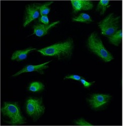
- Experimental details
- Immunofluorescent analysis of GFAP (green) in human diabetic Muller cells. Cells were fixed with 4% paraformaldehyde for 15 minutes, permeabilized with 0.1% Triton X-100 in PBS for 10 minutes at room temperature, and blocked with 5% normal goat serum for 1 hour at room temperature. Cells were probed with a GFAP monoclonal antibody (Product # MA1-35377) at a dilution of 1:25 for 1 hour at room temperature, washed with PBS, and then incubated with an Alexa Fluor 488-conjugated anti-mouse IgG secondary antibody at a dilution of 1:500 for 1 hour at room temperature. Nuclei (blue) were stained with DAPI. Images were taken of a fluorescent microscope at 20X magnification. Data courtesy of the Innovators Program.
