 Explore
 Validate
 Learn
Learn Western blot
 Immunocytochemistry
Antibody data
- Antibody Data
- Antigen structure
- References [1]
- Comments [0]
- Validations
- Western blot [2]
- Immunohistochemistry [8]
- Flow cytometry [2]
Submit
Validation data
Reference
Comment
Report error
- Product number
- NBP2-01761 - Provider product page

- Provider
- Novus Biologicals
- Product name
- Mouse Monoclonal Iduronate 2-Sulfatase/IDS Antibody
- Antibody type
- Monoclonal
- Description
- Affinity purified.
- Reactivity
- Human, Mouse, Rat, Canine, Simian
- Host
- Mouse
- Isotype
- IgG
- Vial size
- 0.1 ml
- Concentration
- 0.71 mg/ml
- Storage
- Store at -20C. Avoid freeze-thaw cycles.
Submitted references FGF signaling deregulation is associated with early developmental skeletal defects in animal models for mucopolysaccharidosis type II (MPSII).
Bellesso S, Salvalaio M, Lualdi S, Tognon E, Costa R, Braghetta P, Giraudo C, Stramare R, Rigon L, Filocamo M, Tomanin R, Moro E
Human molecular genetics 2018 Jul 1;27(13):2262-2275
Human molecular genetics 2018 Jul 1;27(13):2262-2275
No comments: Submit comment
Supportive validation
- Submitted by
- Novus Biologicals (provider)
- Main image

- Experimental details
- Western Blot: Iduronate 2-Sulfatase/IDS Antibody (4G2) [NBP2-01761] - HEK293T cells were transfected with the pCMV6-ENTRY control (Left lane) or pCMV6-ENTRY Iduronate 2 sulfatase (Right lane) cDNA for 48 hrs and lysed. Equivalent amounts of cell lysates (5 ug per lane) were separated by SDS-PAGE and immunoblotted with anti-Iduronate 2 sulfatase.

- Submitted by
- Novus Biologicals (provider)
- Main image

- Experimental details
- Western Blot: Iduronate 2-Sulfatase/IDS Antibody (4G2) [NBP2-01761] - Analysis of extracts (35ug) from 9 different cell lines by using anti-Iduronate 2 sulfatase monoclonal antibody.

Supportive validation
- Submitted by
- Novus Biologicals (provider)
- Main image

- Experimental details
- Immunohistochemistry-Paraffin: Iduronate 2-Sulfatase/IDS Antibody (4G2) [NBP2-01761] - Staining of paraffin-embedded Human prostate tissue using anti-Iduronate 2 sulfatase mouse monoclonal antibody.

- Submitted by
- Novus Biologicals (provider)
- Main image

- Experimental details
- Immunohistochemistry-Paraffin: Iduronate 2-Sulfatase/IDS Antibody (4G2) [NBP2-01761] - Staining of paraffin-embedded Human lymphoma tissue using anti-Iduronate 2 sulfatase mouse monoclonal antibody.

- Submitted by
- Novus Biologicals (provider)
- Main image
- Experimental details
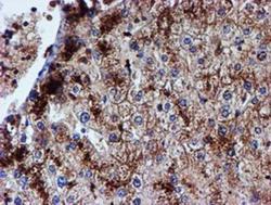
- Immunohistochemistry-Paraffin: Iduronate 2-Sulfatase/IDS Antibody (4G2) [NBP2-01761] - Staining of paraffin-embedded Human liver tissue using anti-Iduronate 2 sulfatase mouse monoclonal antibody.

- Submitted by
- Novus Biologicals (provider)
- Main image

- Experimental details
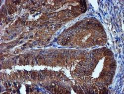
- Immunohistochemistry-Paraffin: Iduronate 2-Sulfatase/IDS Antibody (4G2) [NBP2-01761] - Staining of paraffin-embedded Human Kidney tissue using anti-Iduronate 2 sulfatase mouse monoclonal antibody.

- Submitted by
- Novus Biologicals (provider)
- Main image

- Experimental details
- Immunohistochemistry-Paraffin: Iduronate 2-Sulfatase/IDS Antibody (4G2) [NBP2-01761] - Staining of paraffin-embedded Carcinoma of Human bladder tissue using anti-Iduronate 2 sulfatase mouse monoclonal antibody.

- Submitted by
- Novus Biologicals (provider)
- Main image
- Experimental details
- Immunohistochemistry-Paraffin: Iduronate 2-Sulfatase/IDS Antibody (4G2) [NBP2-01761] - Staining of paraffin-embedded Adenocarcinoma of Human endometrium tissue using anti-Iduronate 2 sulfatase mouse monoclonal antibody.

- Submitted by
- Novus Biologicals (provider)
- Main image

- Experimental details
- Immunohistochemistry-Paraffin: Iduronate 2-Sulfatase/IDS Antibody (4G2) [NBP2-01761] - Human tonsil.

- Submitted by
- Novus Biologicals (provider)
- Main image

- Experimental details
- Immunohistochemistry-Paraffin: Iduronate 2-Sulfatase/IDS Antibody (4G2) [NBP2-01761] - Carcinoma of Human liver tissue.

Supportive validation
- Submitted by
- Novus Biologicals (provider)
- Main image
- Experimental details
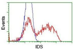
- Flow Cytometry: Iduronate 2-Sulfatase/IDS Antibody (4G2) [NBP2-01761] - HEK293T cells transfected with either overexpression plasmid (Red) or empty vector control plasmid (Blue) were immunostained by anti-Iduronate 2 sulfatase antibody, and then analyzed by flow cytometry.

- Submitted by
- Novus Biologicals (provider)
- Main image
- Experimental details
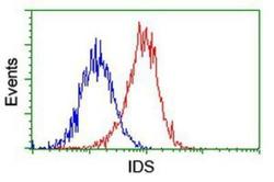
- Flow Cytometry: Iduronate 2-Sulfatase/IDS Antibody (4G2) [NBP2-01761] - Analysis of Hela cells, using anti-Iduronate 2 sulfatase antibody, (Red), compared to a nonspecific negative control antibody (Blue).
