 Explore
 Validate
 Learn
Learn Western blot
 ELISA
ELISAAntibody data
- Antibody Data
- Antigen structure
- References [1]
- Comments [0]
- Validations
- Western blot [2]
- Immunocytochemistry [1]
- Immunohistochemistry [1]
- Other assay [2]
Submit
Validation data
Reference
Comment
Report error
- Product number
- MA5-33115 - Provider product page

- Provider
- Invitrogen Antibodies
- Product name
- ATF2 Recombinant Rabbit Monoclonal Antibody (3D12)
- Antibody type
- Monoclonal
- Antigen
- Synthetic peptide
- Reactivity
- Human, Mouse, Rat
- Host
- Rabbit
- Isotype
- IgG
- Antibody clone number
- 3D12
- Vial size
- 100 µL
- Concentration
- 0.61 mg/mL
- Storage
- -20°C or -80°C if preferred
Submitted references Butorphanol reduces the neuronal inflammatory response and apoptosis via inhibition of p38/JNK/ATF2/p53 signaling.
Huang Y, Li S, Chen H, Feng L, Yuan W, Han T
Experimental and therapeutic medicine 2022 Mar;23(3):229
Experimental and therapeutic medicine 2022 Mar;23(3):229
No comments: Submit comment
Supportive validation
- Submitted by
- Invitrogen Antibodies (provider)
- Main image

- Experimental details
- Western Blot analysis of ATF2 using a ATF2 Monoclonal antibody (Product # MA5-33115) at a concentration of 1.2 µg/mL. Positive WB detected in: K562 whole cell lysate, 293T whole cell lysate. A secondary Goat polyclonal antibody to rabbit IgG was applied at a 1:50,000 dilution. Observed band size: 70 kDa.

- Submitted by
- Invitrogen Antibodies (provider)
- Main image

- Experimental details
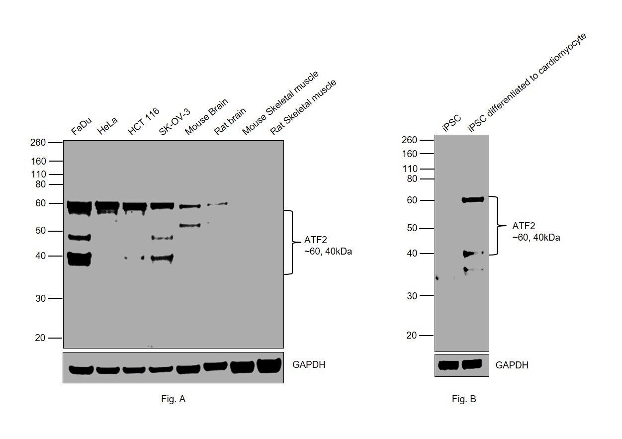
- Western blot was performed using Anti-ATF2 Recombinant Rabbit Monoclonal Antibody (Product # MA5-33115) and a 60 and 40kDa band corresponding to Cyclic AMP-dependent transcription factor ATF-2 was observed across all the tested cell lines and tissues, except Mouse Skeletal Muscle and Rat Skeletal Muscle. Nuclear enriched extracts (30 µg lysate) of FaDu (Fig. A, Lane 1), HeLa (Fig. A, Lane 2), HCT 116 (Fig. A, Lane 3), SK-O-V3 (Fig. A, Lane 4), Mouse Brain (Fig. A, Lane 5), Rat Brain (Fig. A, Lane 6), Mouse Skeletal Muscle (Fig. A, Lane 7), Rat Skeletal Muscle (Fig. A, Lane 8), and Whole cell extracts of iPSC (Fig. B, Lane 1), iPSC (differentiated to cardiomyocytes) (Fig. B, Lane 2) were electrophoresed using NuPAGE™ 10% Bis-Tris Protein Gel (Product # NP0301BOX). Resolved proteins were then transferred onto a Nitrocellulose membrane (Product # IB23001) by iBlot® 2 Dry Blotting System (Product # IB21001). The blot was probed with the primary antibody (1:2000) and detected by chemiluminescence with Goat anti-Rabbit IgG (H+L) Superclonal™ Recombinant Secondary Antibody, HRP (Product # A27036, 1:4000) using the iBright FL 1000 (Product # A32752). Chemiluminescent detection was performed using Novex® ECL Chemiluminescent Substrate Reagent Kit (Product # WP20005).
Supportive validation
- Submitted by
- Invitrogen Antibodies (provider)
- Main image

- Experimental details
- Immunofluorescence analysis of Cyclic AMP-dependent transcription factor ATF-2 was performed using 70% confluent log phase FaDu cells. The cells were fixed with 4% paraformaldehyde for 10 minutes, permeabilized with 0.1% Triton™ X-100 for 15 minutes, and blocked with 2% BSA for 45 minutes at room temperature. The cells were labeled with ATF2 Recombinant Rabbit Monoclonal Antibody (Product # MA5-33115) at 5µg/mL in 0.1% BSA, incubated at 4 degree celsius overnight and then labeled with Goat anti-Rabbit IgG (H+L) Superclonal™ Recombinant Secondary Antibody, Alexa Fluor® 488 conjugate (Product # A27034), (1:2000), for 45 minutes at room temperature (Panel a: Green). Nuclei (Panel b:Blue) were stained with ProLong™ Diamond Antifade Mountant with DAPI (Product # P36962). F-actin (Panel c: Red) was stained with Rhodamine Phalloidin (Product # R415, 1:300). Panel d represents the merged image showing nuclear and cytoplasmic localization. Panel e represents control cells with no primary antibody to assess background. The images were captured at 60X magnification.

Supportive validation
- Submitted by
- Invitrogen Antibodies (provider)
- Main image

- Experimental details
- Immunohistochemical analysis of ATF2 in paraffin embedded human prostate tissue using a ATF2 monoclonal antibody (Product # MA5-33115) at a dilution of 1:115.5. After dewaxing and hydration, antigen retrieval was mediated by high pressure in a citrate buffer (pH 6.0). Section was blocked with 10% normal goat serum 30min at RT. Then primary antibody (1% BSA) was incubated at 4°C overnight. The primary is detected by a biotinylated secondary antibody and visualized using an HRP conjugated SP system.

Supportive validation
- Submitted by
- Invitrogen Antibodies (provider)
- Main image
- Experimental details
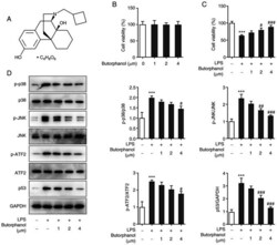
- Effect of butorphanol on LPS-induced decrease in PC12 cell viability and p38/JNK/ATF2/p53 signaling. (A) Chemical structure of butorphanol. (B) The effect of different doses of butorphanol on PC12 cell viability was assessed using a CCK8 assay. (C) The viability of cells treated with butorphanol and LPS was determined using a CCK8 assay. (D) The expression levels of p-p38, p-JNK, p-ATF2, p53, p38, JNK and ATF2 were measured using western blot analysis. *** P

- Submitted by
- Invitrogen Antibodies (provider)
- Main image

- Experimental details
- Butorphanol protects cells against the inhibitory effects of LPS on viability via p38/JNK/ATF2/p53 signaling. (A) The protein expression levels of p-p38, p-JNK, p-ATF2, p53, p38, JNK and ATF2 in each group were determined using western blot analysis. (B) Cell viability in each group was assessed using a CCK8 assay. (C) The activity of LDH in each group was measured using assay kits. ** P
