 Explore
 Validate
 Learn
LearnGTX15731
antibody from GeneTex
Targeting: CREB1
 Western blot
 ELISA Immunocytochemistry Immunoprecipitation Immunohistochemistry Chromatin Immunoprecipitation
Antibody data
- Antibody Data
- Antigen structure
- References [0]
- Comments [0]
- Validations
- Western blot [2]
- Immunocytochemistry [2]
- Immunoprecipitation [1]
- Immunohistochemistry [2]
Submit
Validation data
Reference
Comment
Report error
- Product number
- GTX15731 - Provider product page

- Provider
- GeneTex
- Product name
- CREB antibody [LB9]
- Antibody type
- Monoclonal
- Reactivity
- Human, Mouse, Rat, Simian
- Host
- Mouse
No comments: Submit comment
Supportive validation
- Submitted by
- GeneTex (provider)
- Main image

- Experimental details
- Western blot analysis of CREB in HepG2 cells.
- Submitted by
- GeneTex (provider)
- Main image
- Experimental details
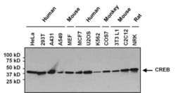
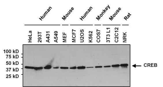
- WB analysis of indicated lysates (25 ug per lane) using CREB antibody [LB9] at a dilution of 1:500.
Supportive validation
- Submitted by
- GeneTex (provider)
- Main image

- Experimental details
- Immunofluorescent analysis of CREB (green) in HeLa cells. Formalin-fixed cells were permeabilized with 0.1% Triton X-100 in TBS for 15 minutes at room temperature. Cells were then blocked with 5% normal goat serum for 15 minutes at room temperature. Cells were probed with CREB antibody [LB9] at a dilution of 1:400 for at least 1 hour at room temperature. Cells were washed with PBS and incubated with DyLight 488-conjugated secondary antibody. Nuclei were stained with Hoechst 33342 dye. Images were taken at 20X magnification.
- Submitted by
- GeneTex (provider)
- Main image

- Experimental details
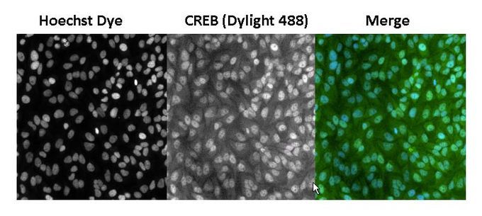
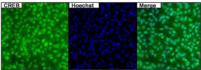
- ICC/IF analysis of HeLa cells using CREB antibody [LB9] at a dilution of 1:500 (green) and Hoechst 33342 dye (blue).
Supportive validation
- Submitted by
- GeneTex (provider)
- Main image

- Experimental details
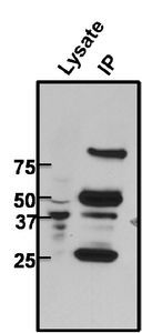
- Immunoprecipitation of CREB from 293T cell lysate. The antigen-antbody complex was formed by binding 500 ug of whole cell lysate with 2 ug of CREB antibody [LB9] overnight on a rocking platform at 4¢XC. The immune-complex was then captured on 50 ul Protein A/G Agarose. Captured immune-complexes were then washed extensively and eluted. Samples were then resolved on a 4-20% Tris-HCl polyacrylamide gel. Proteins were transferred to PVDF membrane and blocked with 5% Milk/TBS-0.1%Tween for at least 1 hour. Membranes were then probed with CREB antibody [LB9] at a dilution of 1:1000 overnight rotating at 4¢XC. Membranes were then washed in TBST and probed with a HRP-conjugated secondary antibody. Membranes were washed and chemiluminescent detection was performed.
Supportive validation
- Submitted by
- GeneTex (provider)
- Main image

- Experimental details
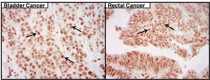
- Immunohistochemistry was performed on cancer biopsies of deparaffinized human bladder and rectal tissue. To expose target proteins high pressure heat induced antigen retrieval was performed using 10mM sodium citrate (pH6.0) buffer for 20 minutes. Following antigen retrieval endogenous peroxidase activity was quenched with 3% hydrogen peroxide for 10 minutes at room temperature. Tissues were then washed in PBS and blocked in 10% normal goat serum for 20 minutes at room temperature. Tissues were probed at a dilution of 1:200 with CREB antibody [LB9] at 4¢XC in a humidified chamber. Tissues were washed extensively with PBS. Colorimetric detection was performed using metal enhanced DAB. Images are displayed at 40X magnification.
- Submitted by
- GeneTex (provider)
- Main image
- Experimental details
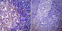
- IHC-P analysis of human tonsil tissue in the presence (left) or abscence (right) of CREB antibody [LB9] at a dilution of 1:100. To expose target proteins, heat induced antigen retrieval was performed using 10mM sodium citrate (pH6.0) buffer, microwaved for 8-15 minutes.
