 Explore
 Validate
 Learn
LearnNB100-74393
antibody from Novus Biologicals
Targeting: CREB1
 Western blot
 ELISA Immunocytochemistry Immunoprecipitation Immunohistochemistry Chromatin Immunoprecipitation
Antibody data
- Antibody Data
- Antigen structure
- References [1]
- Comments [0]
- Validations
- Western blot [2]
- Immunoprecipitation [1]
- Immunohistochemistry [2]
- Chromatin Immunoprecipitation [2]
Submit
Validation data
Reference
Comment
Report error
- Product number
- NB100-74393 - Provider product page

- Provider
- Novus Biologicals
- Proper citation
- Novus Cat#NB100-74393, RRID:AB_1048603
- Product name
- Mouse Monoclonal CREB Antibody
- Antibody type
- Monoclonal
- Description
- Protein A purified. CREB (LB9)
- Reactivity
- Human, Mouse, Rat, Simian
- Host
- Mouse
- Isotype
- IgG
- Vial size
- 100 ug
- Concentration
- 1 mg/ml
- Storage
- Store at -20C. Avoid freeze-thaw cycles.
Submitted references Notch1 Regulates Hippocampal Plasticity Through Interaction with the Reelin Pathway, Glutamatergic Transmission and CREB Signaling.
Brai E, Marathe S, Astori S, Fredj NB, Perry E, Lamy C, Scotti A, Alberi L
Frontiers in cellular neuroscience 2015;9:447
Frontiers in cellular neuroscience 2015;9:447
No comments: Submit comment
Supportive validation
- Submitted by
- Novus Biologicals (provider)
- Main image

- Experimental details
- Western Blot: CREB Antibody (LB9) [NB100-74393] - Proteins were transferred to a PVDF membrane and blocked with 5% Milk/TBST for at least 1 hour. Membranes were probed with a mouse monoclonal antibody recognizing CREB at a dilution of 1:500 overnight at 4C on a rocking platform. Membranes were washed in TBS-0.1%Tween 20 and probed with a goat anti-mouse-HRP secondary antibody at a dilution of 1:20,000 for at least one hour. Membranes were washed and chemiluminescent detection performed.

- Submitted by
- Novus Biologicals (provider)
- Main image

- Experimental details
- Western Blot: CREB Antibody (LB9) [NB100-74393] - Analysis of HepG2 cells

Supportive validation
- Submitted by
- Novus Biologicals (provider)
- Main image
- Experimental details
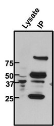
- Immunoprecipitation: CREB Antibody (LB9) [NB100-74393] - Analysis of CREB was performed on untreated 293T cells. Antigen: antbody complex was formed by binding 500ug whole cell lysate with 2ug of mouse monoclonal antibody recognizing CREB overnight on a rocking platform at 4C. The immune-complex was then captured on 50ul Protein A/G Plus Agarose. Captured immune-complexes were then washed extensively and proteins eluted with 5X Reducing Sample Loading Dye. Samples were then resolved on a 4-20% Tris-HCl polyacrylamide gel. Proteins were transferred to PVDF membrane and blocked with 5% Milk/TBS-0.1%Tween for at least 1 hour. Membranes were then probed with a mouse monoclonal antibody recognizing CREB at a dilution of 1:1000 overnight rotating at 4C. Membranes were then washed in TBST and probed with a goat anti-mouse-HRP secondary antibody at a dilution of 1:20,000 for at least one hour. Membranes were washed and chemiluminescent detection was performed using Super Signal West Dura.

Supportive validation
- Submitted by
- Novus Biologicals (provider)
- Main image

- Experimental details
- Immunohistochemistry-Paraffin: CREB Antibody (LB9) [NB100-74393] - Normal biopsies of deparaffinized Human tonsil tissue.

- Submitted by
- Novus Biologicals (provider)
- Main image
- Experimental details
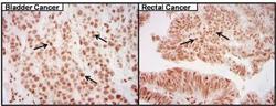
- Immunohistochemistry-Paraffin: CREB Antibody (LB9) [NB100-74393] - Cancer biopsies of deparaffinized human bladder and rectal tissue.

Supportive validation
- Submitted by
- Novus Biologicals (provider)
- Main image

- Experimental details
- Chromatin Immunoprecipitation: CREB Antibody (LB9) [NB100-74393] - Staining of CREB (green) in untreated HeLa cells. Formalin fixed cells were permeabilized with 0.1% Triton X-100 in TBS for 15 minutes at room temperature. Cells were then blocked with 5% normal goat serum for 15 minutes at room temperature. Cells were probed with a mouse monoclonal antibody recognizing CREB at a dilution of 1:400 for at least 1 hour at room temperature. Cells were washed with PBS and incubated with DyLight 488 goat-anti-mouse secondary antibody at a dilution of 1:400 for 30 minutes at room temperature. Nuclei were stained with Hoechst 33342 dye.
- Submitted by
- Novus Biologicals (provider)
- Main image

- Experimental details
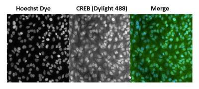
- Chromatin Immunoprecipitation: CREB Antibody (LB9) [NB100-74393] - Analysis of CREB (green) in untreated HeLa cells. Formalin fixed cells were permeabilized with 0.1% Triton X-100 in TBS for 15 minutes at room temperature. Cells were then blocked with 5% normal goat serum for 15 minutes at room temperature. Cells were probed with a mouse monoclonal antibody recognizing CREB at a dilution of 1:500 for at least 1 hour at room temperature. Cells were washed with PBS and incubated with DyLight 488 goat-anti-mouse secondary antibody at a dilution of 1:400 for 30 minutes at room temperature. Nuclei were stained with Hoechst 33342 dye. Images were taken on a Thermo Scientific ArrayScan at 20X magnification.
