 Explore
 Validate
 Learn
Learn Western blot
Antibody data
- Antibody Data
- Antigen structure
- References [1]
- Comments [0]
- Validations
- Western blot [1]
- Immunocytochemistry [2]
- Immunohistochemistry [2]
- Chromatin Immunoprecipitation [1]
Submit
Validation data
Reference
Comment
Report error
- Product number
- AF2989 - Provider product page

- Provider
- R&D Systems
- Product name
- Human CREB Antibody
- Antibody type
- Polyclonal
- Description
- Antigen Affinity-purified. Detects human CREB in Western blots.
- Reactivity
- Human
- Host
- Goat
- Conjugate
- Unconjugated
- Antigen sequence
P16220- Isotype
- IgG
- Vial size
- 100 ug
- Concentration
- LYOPH
- Storage
- Use a manual defrost freezer and avoid repeated freeze-thaw cycles. 12 months from date of receipt, -20 to -70 °C as supplied. 1 month, 2 to 8 °C under sterile conditions after reconstitution. 6 months, -20 to -70 °C under sterile conditions after reconstitution.
Submitted references Dicer1/miR-29/HMGCR axis contributes to hepatic free cholesterol accumulation in mouse non-alcoholic steatohepatitis.
Liu MX, Gao M, Li CZ, Yu CZ, Yan H, Peng C, Li Y, Li CG, Ma ZL, Zhao Y, Pu MF, Miao LL, Qi XM, Ren J
Acta pharmacologica Sinica 2017 May;38(5):660-671
Acta pharmacologica Sinica 2017 May;38(5):660-671
No comments: Submit comment
Supportive validation
- Submitted by
- R&D Systems (provider)
- Main image
- Experimental details
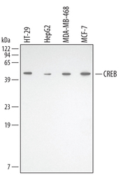
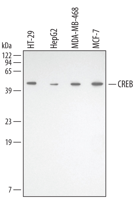
- Detection of Human CREB by Western Blot. Western blot shows lysates of HT-29 human colon adenocarcinoma cell line, HepG2 human hepatocellular carcinoma cell line, MBA-MB-468 human breast cancer cell line, and MCF-7 human breast cancer cell line. PVDF membrane was probed with 0.5 µg/mL of Goat Anti-Human CREB Antigen Affinity-purified Polyclonal Antibody (Catalog # AF2989) followed by HRP-conjugated Anti-Goat IgG Secondary Antibody (Catalog # HAF109). A specific band was detected for CREB at approximately 43 kDa (as indicated). This experiment was conducted under reducing conditions and using Immunoblot Buffer Group 1.
Supportive validation
- Submitted by
- R&D Systems (provider)
- Main image
- Experimental details
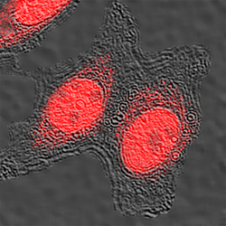
- CREB in MCF-7 Human Cell Line. CREB was detected in immersion fixed MCF-7 human breast cancer cell line using Goat Anti-Human CREB Antigen Affinity-purified Polyclonal Antibody (Catalog # AF2989) at 0.5 µg/mL for 3 hours at room temperature. Cells were stained using the NorthernLights™ 557-conjugated Anti-Goat IgG Secondary Antibody (red; Catalog # NL001). Specific staining was localized to nuclei and cytoplasm. View our protocol for Fluorescent ICC Staining of Cells on Coverslips.

- Submitted by
- R&D Systems (provider)
- Main image

- Experimental details
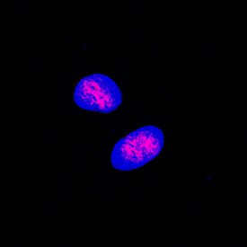
- CREB in MCF-7 Human Cell Line. CREB was detected in immersion fixed MCF-7 human breast cancer cell line using Goat Anti-Human CREB Antigen Affinity-purified Polyclonal Antibody (Catalog # AF2989) at 5 µg/mL for 3 hours at room temperature. Cells were stained using the NorthernLights™ 557-conjugated Anti-Goat IgG Secondary Antibody (red; Catalog # NL001) and counterstained with DAPI (blue). Specific staining was localized to nuclei. View our protocol for Fluorescent ICC Staining of Cells on Coverslips.
Supportive validation
- Submitted by
- R&D Systems (provider)
- Main image
- Experimental details
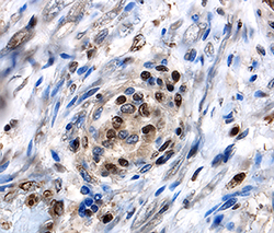
- CREB in Human Dorsal Root Ganglion. CREB was detected in immersion fixed paraffin-embedded sections of human dorsal root ganglion using Goat Anti-Human CREB Antigen Affinity-purified Polyclonal Antibody (Catalog # AF2989) at 10 µg/mL overnight at 4 °C. Before incubation with the primary antibody tissue was subjected to heat-induced epitope retrieval using Antigen Retrieval Reagent-Basic (Catalog # CTS013). Tissue was stained using the Anti-Goat HRP-DAB Cell & Tissue Staining Kit (brown; Catalog # CTS008) and counterstained with hematoxylin (blue). View our protocol for Chromogenic IHC Staining of Paraffin-embedded Tissue Sections.

- Submitted by
- R&D Systems (provider)
- Main image

- Experimental details
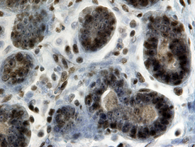
- CREB in Human Breast. CREB was detected in immersion fixed paraffin-embedded sections of human breast using 10 µg/mL Goat Anti-Human CREB Antigen Affinity-purified Polyclonal Antibody (Catalog # AF2989) overnight at 4 °C. Tissue was stained with the Anti-Goat HRP-DAB Cell & Tissue Staining Kit (brown; Catalog # CTS008) and counterstained with hematoxylin (blue). Specific labeling was localized to the nucleus in stromal cells. View our protocol for Chromogenic IHC Staining of Paraffin-embedded Tissue Sections.
Supportive validation
- Submitted by
- R&D Systems (provider)
- Main image
- Experimental details
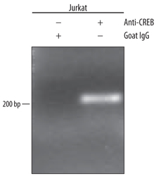
- Detection of CREB-regulated Genes by Chromatin Immunoprecipitation. Jurkat human acute T cell leukemia cell line treated with 50 ng/mL PMA and 200 ng/mL calcium ionomycin for 30 minutes was fixed using formaldehyde, resuspended in lysis buffer, and sonicated to shear chromatin. CREB/DNA complexes were immunoprecipitated using 5 μg Goat Anti-Human CREB Antigen Affinity-purified Polyclonal Antibody (Catalog # AF2989) or control antibody (Catalog # AB-108-C) for 15 minutes in an ultrasonic bath, followed by Biotinylated Anti-Goat IgG Secondary Antibody (Catalog # BAF109). Immunocomplexes were captured using 50 μL of MagCellect Streptavidin Ferrofluid (Catalog # MAG999) and DNA was purified using chelating resin solution. The fos promoter was detected by standard PCR.
