Explore
 Validate
 Learn
Learn Western blot
Antibody data
- Antibody Data
- Antigen structure
- References [21]
- Comments [0]
- Validations
- Western blot [4]
- Immunocytochemistry [2]
- Immunohistochemistry [1]
- Other assay [3]
Submit
Validation data
Reference
Comment
Report error
- Product number
- MA5-13356 - Provider product page

- Provider
- Invitrogen Antibodies
- Product name
- Paxillin Monoclonal Antibody (5H11)
- Antibody type
- Monoclonal
- Antigen
- Purifed from natural sources
- Description
- MA5-13356 targets Paxillin in IF, IHC (P), IP, and WB applications and shows reactivity with Human and Rat samples.
- Antibody clone number
- 5H11
- Concentration
- 0.2 mg/mL
Submitted references Fibroblast growth factor 2 inhibits myofibroblastic activation of valvular interstitial cells.
Topography-induced large-scale antiparallel collective migration in vascular endothelium.
Label2label: training a neural network to selectively restore cellular structures in fluorescence microscopy.
Coordination of mitochondrial and cellular dynamics by the actin-based motor Myo19.
HDAC6 regulates microtubule stability and clustering of AChRs at neuromuscular junctions.
GPR124 facilitates pericyte polarization and migration by regulating the formation of filopodia during ischemic injury.
Expression of claudin, paxillin and FRA-1 in non-nodular breast lesions in association with microcalcifications.
Targeting focal adhesion turnover in invasive breast cancer cells by the purine derivative reversine.
Decreased ezrin and paxillin expression in human urothelial bladder tumors correlate with tumor progression.
Fascin-1, ezrin and paxillin contribute to the malignant progression and are predictors of clinical prognosis in laryngeal squamous cell carcinoma.
The influence of tubular phenotypic changes on the development of diffuse interstitial fibrosis in renal allografts.
Paxillin expression and amplification in early lung lesions of high-risk patients, lung adenocarcinoma and metastatic disease.
Focal adhesion plaque associated cytoskeletons are involved in the invasion and metastasis of human colorectal carcinoma.
Extraribosomal function of metallopanstimulin-1: reducing paxillin in head and neck squamous cell carcinoma and inhibiting tumor growth.
Cyclic strain modulates migration and proliferation of vascular smooth muscle cells via Rho-GDIalpha, Rac1, and p38 pathway.
Vascular smooth muscle cells promote endothelial cell adhesion via microtubule dynamics and activation of paxillin and the extracellular signal-regulated kinase (ERK) pathway in a co-culture system.
Presenilin 1 affects focal adhesion site formation and cell force generation via c-Src transcriptional and posttranslational regulation.
Paxillin is a target for somatic mutations in lung cancer: implications for cell growth and invasion.
The expression of the cytoskeletal focal adhesion protein paxillin in breast cancer correlates with HER2 overexpression and may help predict response to chemotherapy: a retrospective immunohistochemical study.
Smooth muscle alpha-actin deficiency in myofibroblasts leads to enhanced renal tissue fibrosis.
Sequential activation of RhoA and FAK/paxillin leads to ATP release and actin reorganization in human endothelium.
Ground M, Waqanivavalagi S, Park YE, Callon K, Walker R, Milsom P, Cornish J
PloS one 2022;17(6):e0270227
PloS one 2022;17(6):e0270227
Topography-induced large-scale antiparallel collective migration in vascular endothelium.
Leclech C, Gonzalez-Rodriguez D, Villedieu A, Lok T, Déplanche AM, Barakat AI
Nature communications 2022 May 19;13(1):2797
Nature communications 2022 May 19;13(1):2797
Label2label: training a neural network to selectively restore cellular structures in fluorescence microscopy.
Kölln LS, Salem O, Valli J, Hansen CG, McConnell G
Journal of cell science 2022 Feb 1;135(3)
Journal of cell science 2022 Feb 1;135(3)
Coordination of mitochondrial and cellular dynamics by the actin-based motor Myo19.
Majstrowicz K, Honnert U, Nikolaus P, Schwarz V, Oeding SJ, Hemkemeyer SA, Bähler M
Journal of cell science 2021 May 15;134(10)
Journal of cell science 2021 May 15;134(10)
HDAC6 regulates microtubule stability and clustering of AChRs at neuromuscular junctions.
Osseni A, Ravel-Chapuis A, Thomas JL, Gache V, Schaeffer L, Jasmin BJ
The Journal of cell biology 2020 Aug 3;219(8)
The Journal of cell biology 2020 Aug 3;219(8)
GPR124 facilitates pericyte polarization and migration by regulating the formation of filopodia during ischemic injury.
Chen DY, Sun NH, Lu YP, Hong LJ, Cui TT, Wang CK, Chen XH, Wang SS, Feng LL, Shi WX, Fukunaga K, Chen Z, Lu YM, Han F
Theranostics 2019;9(20):5937-5955
Theranostics 2019;9(20):5937-5955
Expression of claudin, paxillin and FRA-1 in non-nodular breast lesions in association with microcalcifications.
Kandelman JD, Waitzberg AF, Szejnfeld J, Smith RL
Sao Paulo medical journal = Revista paulista de medicina 2013;131(2):71-9
Sao Paulo medical journal = Revista paulista de medicina 2013;131(2):71-9
Targeting focal adhesion turnover in invasive breast cancer cells by the purine derivative reversine.
Bijian K, Lougheed C, Su J, Xu B, Yu H, Wu JH, Riccio K, Alaoui-Jamali MA
British journal of cancer 2013 Nov 26;109(11):2810-8
British journal of cancer 2013 Nov 26;109(11):2810-8
Decreased ezrin and paxillin expression in human urothelial bladder tumors correlate with tumor progression.
Athanasopoulou A, Aroukatos P, Nakas D, Repanti M, Papadaki H, Bravou V
Urologic oncology 2013 Aug;31(6):836-42
Urologic oncology 2013 Aug;31(6):836-42
Fascin-1, ezrin and paxillin contribute to the malignant progression and are predictors of clinical prognosis in laryngeal squamous cell carcinoma.
Gao W, Zhang C, Feng Y, Chen G, Wen S, Huangfu H, Wang B
PloS one 2012;7(11):e50710
PloS one 2012;7(11):e50710
The influence of tubular phenotypic changes on the development of diffuse interstitial fibrosis in renal allografts.
Özdemir BH, Özdemir AA, Colak T, Sezer S, Haberal M
Transplantation proceedings 2011 Mar;43(2):527-9
Transplantation proceedings 2011 Mar;43(2):527-9
Paxillin expression and amplification in early lung lesions of high-risk patients, lung adenocarcinoma and metastatic disease.
Mackinnon AC, Tretiakova M, Henderson L, Mehta RG, Yan BC, Joseph L, Krausz T, Husain AN, Reid ME, Salgia R
Journal of clinical pathology 2011 Jan;64(1):16-24
Journal of clinical pathology 2011 Jan;64(1):16-24
Focal adhesion plaque associated cytoskeletons are involved in the invasion and metastasis of human colorectal carcinoma.
Yang HJ, Chen JZ, Zhang WL, Ding YQ
Cancer investigation 2010 Feb;28(2):127-34
Cancer investigation 2010 Feb;28(2):127-34
Extraribosomal function of metallopanstimulin-1: reducing paxillin in head and neck squamous cell carcinoma and inhibiting tumor growth.
Dai Y, Pierson SE, Dudney WC, Stack BC Jr
International journal of cancer 2010 Feb 1;126(3):611-9
International journal of cancer 2010 Feb 1;126(3):611-9
Cyclic strain modulates migration and proliferation of vascular smooth muscle cells via Rho-GDIalpha, Rac1, and p38 pathway.
Qi YX, Qu MJ, Yan ZQ, Zhao D, Jiang XH, Shen BR, Jiang ZL
Journal of cellular biochemistry 2010 Apr 1;109(5):906-14
Journal of cellular biochemistry 2010 Apr 1;109(5):906-14
Vascular smooth muscle cells promote endothelial cell adhesion via microtubule dynamics and activation of paxillin and the extracellular signal-regulated kinase (ERK) pathway in a co-culture system.
Wang YH, Yan ZQ, Shen BR, Zhang L, Zhang P, Jiang ZL
European journal of cell biology 2009 Nov;88(11):701-9
European journal of cell biology 2009 Nov;88(11):701-9
Presenilin 1 affects focal adhesion site formation and cell force generation via c-Src transcriptional and posttranslational regulation.
Waschbüsch D, Born S, Niediek V, Kirchgessner N, Tamboli IY, Walter J, Merkel R, Hoffmann B
The Journal of biological chemistry 2009 Apr 10;284(15):10138-49
The Journal of biological chemistry 2009 Apr 10;284(15):10138-49
Paxillin is a target for somatic mutations in lung cancer: implications for cell growth and invasion.
Jagadeeswaran R, Surawska H, Krishnaswamy S, Janamanchi V, Mackinnon AC, Seiwert TY, Loganathan S, Kanteti R, Reichman T, Nallasura V, Schwartz S, Faoro L, Wang YC, Girard L, Tretiakova MS, Ahmed S, Zumba O, Soulii L, Bindokas VP, Szeto LL, Gordon GJ, Bueno R, Sugarbaker D, Lingen MW, Sattler M, Krausz T, Vigneswaran W, Natarajan V, Minna J, Vokes EE, Ferguson MK, Husain AN, Salgia R
Cancer research 2008 Jan 1;68(1):132-42
Cancer research 2008 Jan 1;68(1):132-42
The expression of the cytoskeletal focal adhesion protein paxillin in breast cancer correlates with HER2 overexpression and may help predict response to chemotherapy: a retrospective immunohistochemical study.
Short SM, Yoder BJ, Tarr SM, Prescott NL, Laniauskas S, Coleman KA, Downs-Kelly E, Pettay JD, Choueiri TK, Crowe JP, Tubbs RR, Budd TG, Hicks DG
The breast journal 2007 Mar-Apr;13(2):130-9
The breast journal 2007 Mar-Apr;13(2):130-9
Smooth muscle alpha-actin deficiency in myofibroblasts leads to enhanced renal tissue fibrosis.
Takeji M, Moriyama T, Oseto S, Kawada N, Hori M, Imai E, Miwa T
The Journal of biological chemistry 2006 Dec 29;281(52):40193-200
The Journal of biological chemistry 2006 Dec 29;281(52):40193-200
Sequential activation of RhoA and FAK/paxillin leads to ATP release and actin reorganization in human endothelium.
Hirakawa M, Oike M, Karashima Y, Ito Y
The Journal of physiology 2004 Jul 15;558(Pt 2):479-88
The Journal of physiology 2004 Jul 15;558(Pt 2):479-88
No comments: Submit comment
Supportive validation
- Submitted by
- Invitrogen Antibodies (provider)
- Main image

- Experimental details
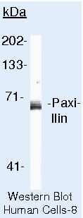
- Western blot of Paxillin using Paxillin Monoclonal Antibody (Product # MA5-13356) on HeLa Cells.
- Submitted by
- Invitrogen Antibodies (provider)
- Main image

- Experimental details
- Western blot analysis was performed on whole cell extracts (30 µg lysate) of A-431 (Lane 1), HeLa (Lane 2), COS-7 (Lane 3), NIH/3T3 (Lane 4), K-562 (Lane 5), Caco-2 (Lane 6), MCF7 (Lane 7), T-47D (Lane 8), A549 (Lane 9) and tissue extract (30 µg lysate) of Mouse Lung (Lane 10). The blots were probed with Anti-Paxillin Monoclonal Antibody (Product # MA5-13356, 2 µg/mL) and detected by chemiluminescence using Goat anti-Mouse IgG (H+L) Superclonal™ Secondary Antibody, HRP conjugate (Product # A28177, 0.25 µg/mL, 1:4000 dilution). A 68 kDa band corresponding to Paxillin was observed across the cell lines and tissue tested. Known quantity of protein samples were electrophoresed using Novex® NuPAGE® 4-12 % Bis-Tris gel (Product # NP0321BOX), XCell SureLock™ Electrophoresis System (Product # EI0002) and Novex® Sharp Pre-Stained Protein Standard (Product # LC5800). Resolved proteins were then transferred onto a nitrocellulose membrane with iBlot® 2 Dry Blotting System (Product # IB21001). The membrane was probed with the relevant primary antibody following blocking with 5 % skimmed milk. Chemiluminescent detection was performed using Pierce™ ECL Western Blotting Substrate (Product # 32106).

- Submitted by
- Invitrogen Antibodies (provider)
- Main image

- Experimental details
- Knockdown of Paxillin was achieved by transfecting HeLa cells with Paxillin specific siRNAs (Silencer® select Product # s11629 and s11627). Western blot analysis (Fig a) was performed using Whole cell extract from the Paxillin knock down cells (lane 3), non-specific scrambled siRNA transfected cells (lane 2) and untransfected cells (lane 1). The blots were probed with Anti-Paxillin Mouse Monoclonal Antibody (Product # MA5-13356, 1 µg/mL) and Goat anti-Mouse IgG (H+L) Superclonal™ Secondary Antibody, HRP conjugate (Product # A28177, 0.25 µg/mL, 1:4000 dilution). Densitometric analysis of this western blot is shown in histogram (Fig b). Loss of signal upon siRNA mediated knock down confirms that antibody is specific to Paxillin.

- Submitted by
- Invitrogen Antibodies (provider)
- Main image

- Experimental details
- Knockout of Paxillin was achieved by CRISPR-Cas9 genome editing using LentiArray™ Lentiviral sgRNA (Product # A32042, Assay ID CRISPR1071991_LV) and LentiArray Cas9 Lentivirus (Product # A32064). Western blot analysis of Paxillin was performed by loading 30 µg of HeLa Wild Type (Lane 1), HeLa Cas9 (Lane 2) andHeLa Paxillin KO (Lane 3) whole cell extracts. The samples were electrophoresed using NuPAGE™ Novex™ 4-12% Bis-Tris Protein Gel (Product # NP0322BOX). Resolved proteins were then transferred onto a nitrocellulose membrane (Product # IB23001) by iBlot® 2 Dry Blotting System (Product # IB21001). The blot was probed with Anti-Paxillin Monoclonal Antibody (5H11) (Product # MA5-13356, 1 µg/mL dilution) and Goat anti-Mouse IgG (H+L) Superclonal™ Recombinant Secondary Antibody, HRP (Product # A28177, 1:5,000 dilution) using the iBright FL 1000 (Product # A32752). Chemiluminescent detection was performed using Novex® ECL Chemiluminescent Substrate Reagent Kit (Product # WP20005). Loss of signal upon CRISPR mediated knockout (KO) using the LentiArray™ CRISPR product line confirms that antibody is specific to Paxillin.

Supportive validation
- Submitted by
- Invitrogen Antibodies (provider)
- Main image

- Experimental details
- Immunofluorescence analysis of Paxillin was performed using 70% confluent log phase HeLa cells. The cells were fixed with 4% paraformaldehyde for 10 minutes, permeabilized with 0.1% Triton™ X-100 for 10 minutes, and blocked with 1% BSA for 1 hour at room temperature. The cells were labeled with Paxillin Monoclonal Antibody (Product # MA5-13356) at 5 µg/mL in 0.1% BSA and incubated for 3 hours at room temperature and then labeled with Goat anti-Mouse IgG (H+L) Superclonal™ Secondary Antibody, Alexa Fluor® 488 conjugate (Product # A28175) at a dilution of 1:2000 for 45 minutes at room temperature (Panel a: green). Nuclei (Panel b: blue) were stained with SlowFade® Gold Antifade Mountant with DAPI (Product # S36938). F-actin (Panel c: red) was stained with Rhodamine Phalloidin (Product # R415, 1:300). Panel d represents the merged image showing cytoplasmic localization. Panel e shows the no primary antibody control. The images were captured at 60X magnification.

- Submitted by
- Invitrogen Antibodies (provider)
- Main image

- Experimental details
- Knockdown of Paxillin was achieved by transfecting HeLa cells with Paxillin specific siRNA (Silencer® select Cat # s11629, s11627). Immunofluorescence analysis was performed on HeLa cells (untransfected, panel a,d), transfected with non-specific scrambled siRNA (panels b,e) and transfected with Paxillin specific siRNA (panel c,f) Cells were fixed, permeabilized, and labelled with Paxillin Mouse monoclonal Antibody (Product # MA5-13356, 5 µg/mL), followed by Goat anti-Mouse IgG (H+L) Superclonal™ Secondary Antibody, Alexa Fluor® 488 conjugate (Product # A28175, 1:2000). Nuclei (blue) were stained using SlowFade® Gold Antifade Mountant with DAPI (Product # S36938), and Rhodamine Phalloidin (Product # R415, 1:300) was used for cytoskeletal F-actin (red) staining. Loss of specific signal was observed upon siRNA mediated knockdown (panel c,f) confirming specificity of the antibody to Paxillin(green). The images were captured at 60X magnification.

Supportive validation
- Submitted by
- Invitrogen Antibodies (provider)
- Main image

- Experimental details
- Formalin-fixed, paraffin-embedded human colon carcinoma stained with Paxillin antibody using peroxidase-conjugate and AEC chromogen. Note cytoplasmic staining of tumor cells.

Supportive validation
- Submitted by
- Invitrogen Antibodies (provider)
- Main image

- Experimental details
- Immunoprecipitation of Paxillin using Paxillin Monoclonal Antibody (Product # MA5-13356) on Native Human A431 Cells.

- Submitted by
- Invitrogen Antibodies (provider)
- Main image

- Experimental details
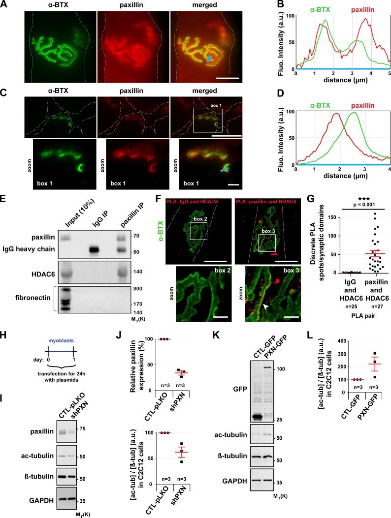
- Figure 5. In muscle cells, paxillin is present at the NMJ and promotes the regulation of MT acetylation. (A and C) Isolated fibers (A) and cross sections (C) of TA muscles from 2-mo-old mice were double-stained with an antibody against paxillin (in red) and with alpha-BTX-A488 (in green). (B and D) The fluorescence intensity of each staining was plotted as a function of the distance (based on the blue line scans in A and C, respectively). The green curve corresponds to alpha-BTX-A488 staining and the red curve to paxillin staining. (E) Western blot showing the co-immunoprecipitation of endogenous HDAC6 and paxillin in TA muscle cells. (F and G) Representative images (F) and quantitation (G) of a PLA performed in isolated fibers of TA muscle with protein-specific antibody pairs as indicated. Cells were counterstained with alpha-BTX-A488 in green ( n = number of synaptic domains quantified). PLA-positive spots are shown in red. The arrowhead shows the colocalization of alpha-BTX and PLA. Means +- SEM. ***, P < 0.001; Mann-Whitney U test. (H) Schematic representation of the experimental time course. (I-L) Myoblasts were transfected with either shRNA-Control (pLKO), shRNA against paxillin (shPXN), GFP alone, or PXN-GFP for 24 h. (I and K) Representative Western blots showing endogenous paxillin, acetylated tubulin (ac-tubulin), GFP, and beta-tubulin expression. GAPDH was used as a loading control. (J and L) Quantification of acetylated tubulin protein level, normalized to beta-tu
- Submitted by
- Invitrogen Antibodies (provider)
- Main image

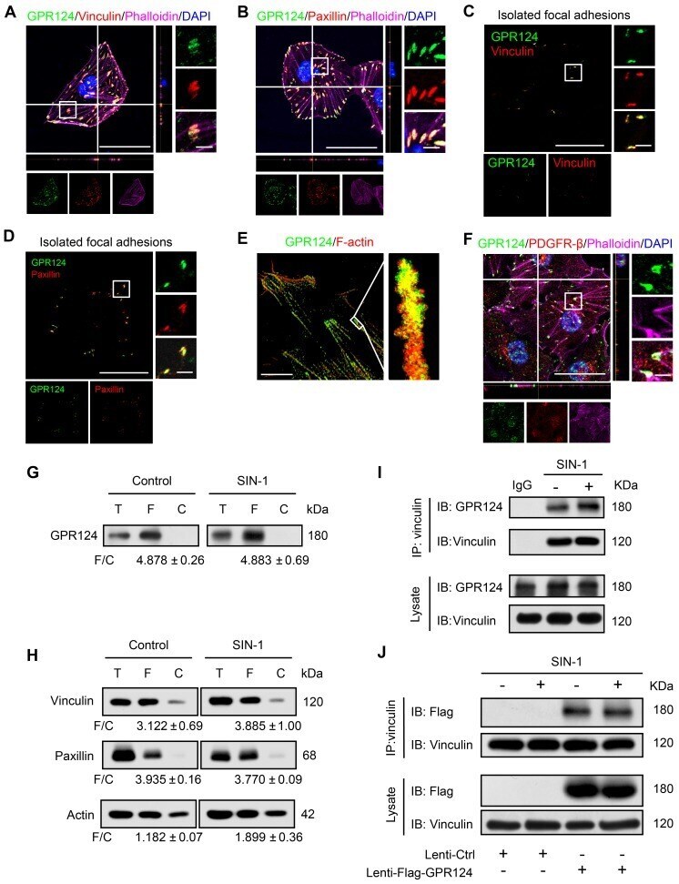
- Experimental details
- Figure 2 GPR124 expression in focal adhesions in HBVPs. ( A, B ) Maximum intensity projections of confocal images from HBVPs stained for GPR124 (green), Vinculin (red, A ), Paxillin (red, B ), Phalloidin (magenta) and DAPI (blue, nuclei). The images on the right panel are magnifications of the boxed regions in the images. Scale bar: 40 mum (Left), magnified images, 5 mum. Representative stainings from three independent experiments are shown. ( C, D ) Isolated focal adhesions include the same proteins as focal adhesions found in intact HBVPs. Confocal fluorescence microscopy images of immunostained isolated focal adhesions demonstrating the presence of GPR124 (green), Vinculin (red, C ) and Paxillin (red, D ). Scale bar: 40 mum (Left), magnified images, 5 mum. ( E ) The localization of GPR124 and F-actin in HBVPs was observed by STORM. Note that there is a close interaction between GPR124 and the F-actin terminals. Scale bar: 10 mum. ( F ) Maximum intensity projections of confocal images from primary mouse brain pericytes stained for GPR124 (green), PDGF-beta (red), Phalloidin (magenta) and DAPI (blue, nuclei). The images on the right panel are magnifications of the boxed regions in the images. Scale bar: 40 mum (Left), magnified images, 5 mum. ( G, H ) Western blot comparison of protein concentration in total cell lysate (T), isolated focal adhesion fractions (F) and cell body fractions (C). Equal total protein was loaded in each lane. The ratio shown on the lower (F/C) indic