Explore
 Validate
 Learn
Learn51-0500
antibody from Invitrogen Antibodies
Targeting: RELA
NFKB3, p65
 Western blot
 ELISA Immunocytochemistry Immunohistochemistry Flow cytometry Chromatin Immunoprecipitation Other assay
Antibody data
- Antibody Data
- Antigen structure
- References [15]
- Comments [0]
- Validations
- Western blot [3]
- Immunocytochemistry [2]
- Flow cytometry [1]
- Chromatin Immunoprecipitation [1]
- Other assay [5]
Submit
Validation data
Reference
Comment
Report error
- Product number
- 51-0500 - Provider product page

- Provider
- Invitrogen Antibodies
- Product name
- NFkB p65 Polyclonal Antibody
- Antibody type
- Polyclonal
- Antigen
- Recombinant full-length protein
- Reactivity
- Human, Mouse
- Host
- Rabbit
- Isotype
- IgG
- Vial size
- 100 µg
- Concentration
- 0.25 mg/mL
- Storage
- -20°C
Submitted references Mechanical Study of Jian-Gan-Xiao-Zhi Decoction on Nonalcoholic Fatty Liver Disease Based on Integrated Network Pharmacology and Untargeted Metabolomics.
β-Nicotinamide Mononucleotide (NMN) Administrated by Intraperitoneal Injection Mediates Protection Against UVB-Induced Skin Damage in Mice.
Fetuin-A secretion from β-cells leads to accumulation of macrophages in islets, aggravates inflammation and impairs insulin secretion.
ECRG4 Represses Cell Proliferation and Invasiveness via NFIC/OGN/NF-κB Signaling Pathway in Bladder Cancer.
Chick Embryo: A Preclinical Model for Understanding Ischemia-Reperfusion Mechanism.
Establishment of three novel cell lines derived from African American patients with colorectal carcinoma: A unique tool for assessing racial health disparity.
Counteraction of Apoptotic and Inflammatory Effects of Adriamycin in the Liver Cell Culture by Clinopitolite.
LPS-induced cytokine levels are repressed by elevated expression of HSP70 in rats: possible role of NF-kappaB.
Promoter targeted small RNAs induce long-term transcriptional gene silencing in human cells.
Expression of angiogenic factors vascular endothelial growth factor and interleukin-8/CXCL8 is highly responsive to ambient glutamine availability: role of nuclear factor-kappaB and activating protein-1.
NF-kappa B in the vascular progression of melanoma.
PKC zeta participates in activation of inflammatory response induced by enteropathogenic E. coli.
Melatonin reduces renal interstitial inflammation and improves hypertension in spontaneously hypertensive rats.
Protection against hypoxia-induced increase in blood-brain barrier permeability: role of tight junction proteins and NFkappaB.
Protection against hypoxia-induced increase in blood-brain barrier permeability: role of tight junction proteins and NFkappaB.
Cao YJ, Li HZ, Zhao J, Sun YM, Jin XW, Lv SQ, Luo JY, Fang XX, Wen WB, Liao JB
Evidence-based complementary and alternative medicine : eCAM 2022;2022:2264394
Evidence-based complementary and alternative medicine : eCAM 2022;2022:2264394
β-Nicotinamide Mononucleotide (NMN) Administrated by Intraperitoneal Injection Mediates Protection Against UVB-Induced Skin Damage in Mice.
Zhou X, Du HH, Long X, Pan Y, Hu J, Yu J, Zhao X
Journal of inflammation research 2021;14:5165-5182
Journal of inflammation research 2021;14:5165-5182
Fetuin-A secretion from β-cells leads to accumulation of macrophages in islets, aggravates inflammation and impairs insulin secretion.
Mukhuty A, Fouzder C, Kundu R
Journal of cell science 2021 Nov 1;134(21)
Journal of cell science 2021 Nov 1;134(21)
ECRG4 Represses Cell Proliferation and Invasiveness via NFIC/OGN/NF-κB Signaling Pathway in Bladder Cancer.
Liang X, Gao J, Wang Q, Hou S, Wu C
Frontiers in genetics 2020;11:846
Frontiers in genetics 2020;11:846
Chick Embryo: A Preclinical Model for Understanding Ischemia-Reperfusion Mechanism.
Fauzia E, Barbhuyan TK, Shrivastava AK, Kumar M, Garg P, Khan MA, Robertson AAB, Raza SS
Frontiers in pharmacology 2018;9:1034
Frontiers in pharmacology 2018;9:1034
Establishment of three novel cell lines derived from African American patients with colorectal carcinoma: A unique tool for assessing racial health disparity.
Paredes J, Ji P, Lacomb JF, Shroyer KR, Martello LA, Williams JL
International journal of oncology 2018 Oct;53(4):1516-1528
International journal of oncology 2018 Oct;53(4):1516-1528
Counteraction of Apoptotic and Inflammatory Effects of Adriamycin in the Liver Cell Culture by Clinopitolite.
Yapislar H, Taskin E, Ozdas S, Akin D, Sonmez E
Biological trace element research 2016 Apr;170(2):373-81
Biological trace element research 2016 Apr;170(2):373-81
LPS-induced cytokine levels are repressed by elevated expression of HSP70 in rats: possible role of NF-kappaB.
Dokladny K, Lobb R, Wharton W, Ma TY, Moseley PL
Cell stress & chaperones 2010 Mar;15(2):153-63
Cell stress & chaperones 2010 Mar;15(2):153-63
Promoter targeted small RNAs induce long-term transcriptional gene silencing in human cells.
Hawkins PG, Santoso S, Adams C, Anest V, Morris KV
Nucleic acids research 2009 May;37(9):2984-95
Nucleic acids research 2009 May;37(9):2984-95
Expression of angiogenic factors vascular endothelial growth factor and interleukin-8/CXCL8 is highly responsive to ambient glutamine availability: role of nuclear factor-kappaB and activating protein-1.
Bobrovnikova-Marjon EV, Marjon PL, Barbash O, Vander Jagt DL, Abcouwer SF
Cancer research 2004 Jul 15;64(14):4858-69
Cancer research 2004 Jul 15;64(14):4858-69
NF-kappa B in the vascular progression of melanoma.
Kashani-Sabet M, Shaikh L, Miller JR 3rd, Nosrati M, Ferreira CM, Debs RJ, Sagebiel RW
Journal of clinical oncology : official journal of the American Society of Clinical Oncology 2004 Feb 15;22(4):617-23
Journal of clinical oncology : official journal of the American Society of Clinical Oncology 2004 Feb 15;22(4):617-23
PKC zeta participates in activation of inflammatory response induced by enteropathogenic E. coli.
Savkovic SD, Koutsouris A, Hecht G
American journal of physiology. Cell physiology 2003 Sep;285(3):C512-21
American journal of physiology. Cell physiology 2003 Sep;285(3):C512-21
Melatonin reduces renal interstitial inflammation and improves hypertension in spontaneously hypertensive rats.
Nava M, Quiroz Y, Vaziri N, Rodriguez-Iturbe B
American journal of physiology. Renal physiology 2003 Mar;284(3):F447-54
American journal of physiology. Renal physiology 2003 Mar;284(3):F447-54
Protection against hypoxia-induced increase in blood-brain barrier permeability: role of tight junction proteins and NFkappaB.
Brown RC, Mark KS, Egleton RD, Huber JD, Burroughs AR, Davis TP
Journal of cell science 2003 Feb 15;116(Pt 4):693-700
Journal of cell science 2003 Feb 15;116(Pt 4):693-700
Protection against hypoxia-induced increase in blood-brain barrier permeability: role of tight junction proteins and NFkappaB.
Brown RC, Mark KS, Egleton RD, Huber JD, Burroughs AR, Davis TP
Journal of cell science 2003 Feb 15;116(Pt 4):693-700
Journal of cell science 2003 Feb 15;116(Pt 4):693-700
No comments: Submit comment
Supportive validation
- Submitted by
- Invitrogen Antibodies (provider)
- Main image

- Experimental details
- Western blot analysis of NFkB (p65) was performed by loading 30 µg of HeLa (lane1), HEK-293 (lane2), A-431 (lane3), K562 (lane4) Cos-7 (lane5), NIH/3T3 (lane 6), MDCK (lane 7) cell lysate using Novex®NuPAGE®4-12 % Bis-Tris gel (Product # NP0321BOX), XCell SureLock Electrophoresis System (Product # EI0002), Novex® Sharp Pre-Stained Protein Standard (LC5800), and iBlot® Dry Blotting System (IB21001). Proteins were transferred to a nitrocellulose membrane and blocked with 5% skim milk for 1 hour at room temperature. NFkB (p65) was detected at ~ 65 kDa using NFkB (p65) Rabbit Polyclonal Antibody (Product # 51-0500) at 1-3 µg/mL in 5% skim milk at 4°C overnight on a rocking platform. Goat Anti-Rabbit IgG - HRP Secondary Antibody (G21234) at 1:5000 dilution was used and chemiluminescent detection was performed using Pierce™ ECL Western Blotting Substrate (Product # 32106).

- Submitted by
- Invitrogen Antibodies (provider)
- Main image

- Experimental details
- Knockdown of NFkB p65 was achieved by transfecting HeLa with NFkB p65 specific siRNAs (Silencer® select Product # s11916, s11914). Western blot analysis was performed using Nuclear enriched extracts from the NFkB p65 knockdown cells (lane 3), scrambled siRNA transfected cells (lane 2), and untransfected cells (lane 1). The blot was probed with NFkB p65 Polyclonal Antibody (Product # 51-0500, 0.25 µg/mL) and Goat anti-Rabbit IgG (H+L) Superclonal™ Recombinant Secondary Antibody, HRP (Product # A27036, 1:20000 dilution). Decrease in signal upon siRNA mediated knock down confirms that antibody is specific to NFkB p65.

Supportive validation
- Submitted by
- Invitrogen Antibodies (provider)
- Main image
- Experimental details
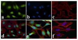
- Immunofluorescence analysis of NFkB (p65) was done on 70% confluent log phase HeLa cells treated with TNF-alpha (50 ng/mL for 20 min). The cells were fixed with 4% paraformaldehyde for 15 minutes, permeabilized with 0.25% Triton X-100 for 10 minutes, and blocked with 5% BSA for 1 hour at room temperature. The cells were labeled with NFkB (p65) Rabbit polyclonal Antibody (Product # 51-0500) at 2 µg/mL in 1% BSA and incubated for 3 hours at room temperature and then labeled with Alexa Fluor 488 Goat Anti-Rabbit IgG Secondary Antibody (Product # A-11008) at a dilution of 1:400 for 30 minutes at room temperature (Panel a: green). Nuclei (Panel b: blue) were stained with SlowFade® Gold Antifade Mountant DAPI (Product # S36938). F-actin (Panel c: red) was stained with Alexa Fluor 594 Phalloidin (Product # A12381). Panel d is a merged image showing nuclear localization upon treatment. Panel e shows untreated HeLa cells with cytoplasmic localization. Panel f shows no primary antibody control. The images were captured at 20X magnification.

- Submitted by
- Invitrogen Antibodies (provider)
- Main image

- Experimental details
- Immunofluorescence analysis of NFkB p65 was done on 70% confluent log phase ME-180 cells. The cells were either mock treated or treated with TNF-alpha (50 ng/mL for 20 min), fixed with 4% paraformaldehyde for 15 minutes, permeabilized with 0.1% Triton™ X-100 for 10 minutes, and blocked with 1% BSA for 1 hour at room temperature. The cells were subsequently labeled with NFkB p65 (Green) Rabbit Polyclonal Antibody (510500) at 1:300 in 0.1% BSA and incubated for 3 hours at room temperature and then labeled with Goat anti-Rabbit IgG (H+L) Superclonal™ Secondary Antibody, Alexa Fluor® 488 conjugate (Product # A27034) at a dilution of 1:2000 for 45 minutes at room temperature. Nuclei (Blue) were stained with SlowFade® Gold Antifade Mountant DAPI (Product # S36938). F-actin (Red) was stained with Rhodamine Phalloidin (Product # R415, 1:300). TNFalpha induced nuclear translocation of NFkB, a downstream target in the TNFR1, TRADD and IKK alpha was observed in control cell line (panels a, e) and not in the TNFR1, TRADD and IKK alpha knockout (KO) cell lines (panels b-d; f-h). The images were captured at 40X magnification.

Supportive validation
- Submitted by
- Invitrogen Antibodies (provider)
- Main image

- Experimental details
- Flow cytometry analysis of NFkB [p65] was done on HeLa cells. Cells were fixed with 70% ethanol for 10 minutes, permeabilized with 0.25% Tritonª X-100 for 20 minutes, and blocked with 5% BSA for 30 minutes at room temperature. Cells were labeled with NFkB [p65] Rabbit Polyclonal Antibody (510500, red histogram) or with rabbit isotype control (pink histogram) at 3-5 µg/million cells in 2.5% BSA. After incubation at room temperature for 2 hours, the cells were labeled with Alexa Fluor¨ 488 Goat Anti-Rabbit Secondary Antibody (A11008) at a dilution of 1:400 for 30 minutes at room temperature. The representative 10,000 cells were acquired and analyzed for each sample using an Attune¨ Acoustic Focusing Cytometer. The purple histogram represents unstained control cells and the green histogram represents no-primary-antibody control.

Supportive validation
- Submitted by
- Invitrogen Antibodies (provider)
- Main image

- Experimental details
- ChIP- qPCR analysis of NFkB p65 was performed with 5 µg of the NFkB p65 Rabbit polyclonal antibody (Product # 51-0500) on sheared chromatin from 2 million HeLa cells treated with 50 ng/mL of TNFalpha for one hour using the MAGnify™ Chromatin Immunoprecipitation System (Product # 49-2024). Normal Rabbit IgG was used as a negative IP control. The purified DNA from each ChIP sample was analyzed by StepOnePlus™ Real-Time PCR System (Product # 4376600) with primers for the promoter of active IL-6, IL-8 gene, used as positive control target, and the inactive GAPDH, used as negative control target. Data is presented as fold enrichment of the antibody signal versus the negative control IgG using the comparative CT method.

Supportive validation
- Submitted by
- Invitrogen Antibodies (provider)
- Main image
- Experimental details
- NULL

- Submitted by
- Invitrogen Antibodies (provider)
- Main image

- Experimental details
- Figure 2. Mechanism of UBC167 induced silencing. ( A ) UBC167-mediated suppression of UbC expression in 293Gt cells is operative via transcriptional gene silencing. Nuclear run-on assay was carried out on UBC167 or Control (CCR5) duplex RNA treated cultures 48 h post-transfection. Run-on RNA samples containing incorporated biotin UTP were immunopreciptated and analyzed by dot blot and qRT-PCR ( 29 ). ( B , C ) UBC167 and UBC308 direct silent state histone marks H3K9me2 and H3K27me3, and Ago-1 to the targeted locus. UBC167, UBC308 or CCR5 (control) dsRNA-treated 293Gt cells were assessed by ChIP for H3K9me2, H3K27me3 and Ago-1 at various regions of the UbC gene. Cultures were assessed 48 h post-dsRNA transfection with primers spanning the target site and downstream of the known TATAA site. Standard errors of the means are shown from quadruplicate duplex RNA treated cultures. ( D ) Activating chromatin marks are lost following UBC167 treatment, while RNAPII remains. ChIP analysis was performed using antibodies against NF-kB (p65), AcH3K14 and RNAPII, followed by PCR and qPCR with UbC promoter-specific primers. Samples were isolated from stable Tet-inducible 293Trex-UBC167 cells treated with Tet for 3 days.

- Submitted by
- Invitrogen Antibodies (provider)
- Main image
- Experimental details
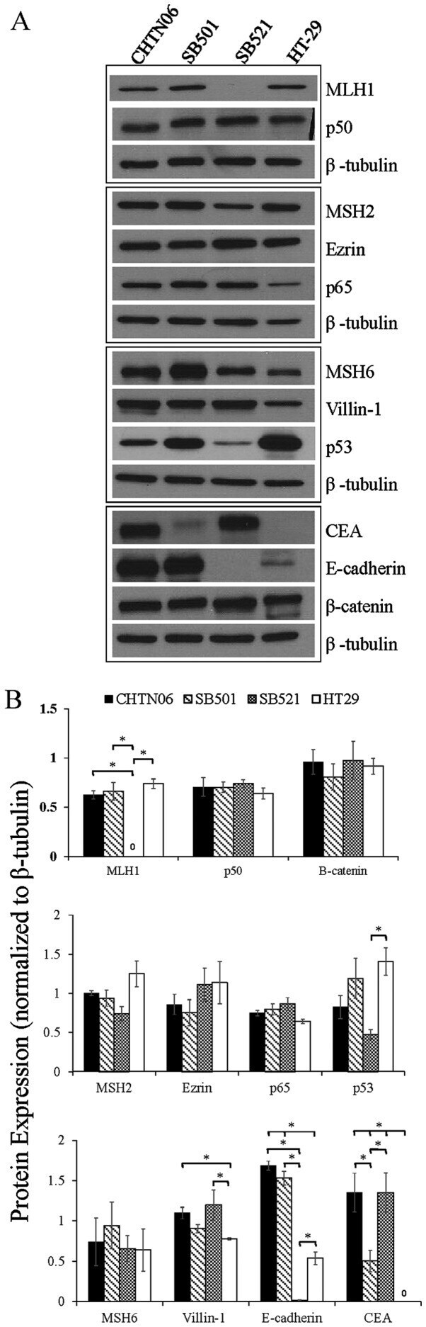
- Figure 3 The expression of proteins associated with colorectal carcinoma (CRC) tumorigenesis and metastasis was determined in the novel African American CRC lines by immunoblotting. (A) Qualitative analysis of CHTN06, SB501 and SB521 and HT-29, a Caucasian American CRC cell line, for protein expression of beta-catenin, p53, nuclear factor (NF)-kappaB (p50 and p65), villin-1, MSH2, MSH6, MLH1 and ezrin. (B) Semi-quantitative densitometry was performed by normalizing protein expression to the respective beta-tubulin loading control. Data were generated from three independent experiments. CEA, carcinoembryonic antigen.
- Submitted by
- Invitrogen Antibodies (provider)
- Main image

- Experimental details
- Figure 6 Protein expression of AMPK, NFkappaB-p65, IkappaB-alpha, SOD1 and CAT in skin tissues. ( A ) relative expression levels of proteins; ( B ) protein banding map. * p < 0.05 compared to the UVB group; ** p < 0.01 compared to the UVB group; *** p < 0.001 compared to the UVB group. Abbreviations : VC+UVB, mice treated with vitamin C(300mg/kg) and UVB irradiation; NMN+UVB, mice treated with nicotinamide mononucleotide (300mg/kg) and UVB irradiation.

- Submitted by
- Invitrogen Antibodies (provider)
- Main image

- Experimental details
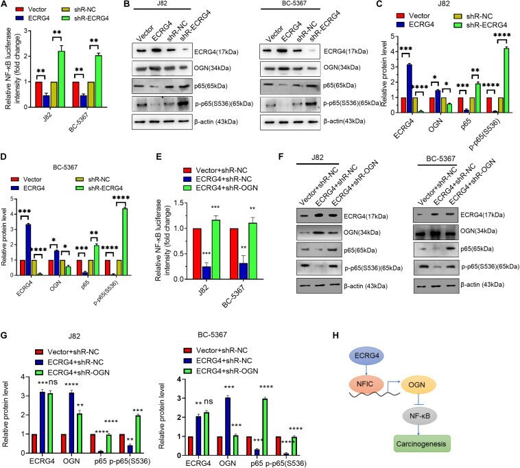
- FIGURE 6 ECRG4 inhibits the NF-kappaB pathway via the upregulation of OGN in BCa cells. (A) NF-kappaB transcriptional activity was detected using an NF-kappaB luciferase reporter gene system. (B) Western blotting detected the protein levels of ECRG4, OGN, p65, and p-p65 (S536) in BCa cells after transfection with ECRG4-overexpressing or silencing plasmids. (C,D) Quantification of the protein levels of ECRG4, OGN, p65, and p-p65 (S536) in panel (B) . (E) The NF-kappaB luciferase reporter gene system was used to detect the NF-kappaB transcriptional activity of BCa cells after transfection with indicated plasmids. (F) Western blotting detected the protein levels of p65 and p-p65 (S536) in BCa cells after transfection with the indicated plasmids. (G) Quantification of the protein levels of p65 and p-p65 (S536) in panel (F) . (H) The proposed mechanism of ECRG4 inhibition of the carcinogenesis of BCa cells via NFIC/OGN/NF-kappaB signaling. (A-G) ANOVA followed by Bonferroni's post hoc test. All data were replicated three times. * p < 0.05, ** p < 0.01, *** p < 0.001, **** p < 0.0001.