 Explore
 Validate
 Learn
Learn Western blot
 Immunoprecipitation
Antibody data
- Antibody Data
- Antigen structure
- References [11]
- Comments [0]
- Validations
- Western blot [4]
- Flow cytometry [1]
- Chromatin Immunoprecipitation [1]
- Other assay [9]
Submit
Validation data
Reference
Comment
Report error
- Product number
- PA1-186 - Provider product page

- Provider
- Invitrogen Antibodies
- Product name
- NFkB p65 Polyclonal Antibody
- Antibody type
- Polyclonal
- Antigen
- Other
- Description
- PA1-186 has been successfully used in WB using StartingBlock T20 (TBS) Blocking Buffer (Product # 37543). More non-specific bands are observed when using 5% BSA for blocking. By WB, PA1-186 detects a predominant band at 65kD. Loading increasing amounts of cell lysate may yield more intense nonspecific bands at ~120kD and/or ~50kD. PA1-186 has been succesfully used for IP of the NF-kB p65 subunit and co-IP of the NF-kB p50 subunit.
- Reactivity
- Human, Mouse
- Host
- Rabbit
- Isotype
- IgG
- Vial size
- 100 µg
- Concentration
- 1 mg/mL
- Storage
- -20°C
Submitted references Selective GSK3β Inhibition Mediates an Nrf2-Independent Anti-inflammatory Microglial Response.
SIRT1-Dependent Upregulation of BDNF in Human Microglia Challenged with Aβ: An Early but Transient Response Rescued by Melatonin.
Myeloid Differentiation Primary Response 88-Cyclin D1 Signaling in Breast Cancer Cells Regulates Toll-Like Receptor 3-Mediated Cell Proliferation.
SIRT1 Mediates Melatonin's Effects on Microglial Activation in Hypoxia: In Vitro and In Vivo Evidence.
Short-Term Microgravity Influences Cell Adhesion in Human Breast Cancer Cells.
Polyphenols in Liubao Tea Can Prevent CCl₄-Induced Hepatic Damage in Mice through Its Antioxidant Capacities.
Mitigating SOX2-potentiated Immune Escape of Head and Neck Squamous Cell Carcinoma with a STING-inducing Nanosatellite Vaccine.
CaMKIIα expression in a mouse model of NMDAR hypofunction schizophrenia: Putative roles for IGF-1R and TLR4.
Connexin 43 Controls the Astrocyte Immunoregulatory Phenotype.
Hepatoprotective Effects of Lactobacillus on Carbon Tetrachloride-Induced Acute Liver Injury in Mice.
MicroRNA-27a Inhibits Cell Migration and Invasion of Fibroblast-Like Synoviocytes by Targeting Follistatin-Like Protein 1 in Rheumatoid Arthritis.
Yousef MH, Salama M, El-Fawal HAN, Abdelnaser A
Molecular neurobiology 2022 Sep;59(9):5591-5611
Molecular neurobiology 2022 Sep;59(9):5591-5611
SIRT1-Dependent Upregulation of BDNF in Human Microglia Challenged with Aβ: An Early but Transient Response Rescued by Melatonin.
Caruso GI, Spampinato SF, Costantino G, Merlo S, Sortino MA
Biomedicines 2021 Apr 24;9(5)
Biomedicines 2021 Apr 24;9(5)
Myeloid Differentiation Primary Response 88-Cyclin D1 Signaling in Breast Cancer Cells Regulates Toll-Like Receptor 3-Mediated Cell Proliferation.
Singh A, Devkar R, Basu A
Frontiers in oncology 2020;10:1780
Frontiers in oncology 2020;10:1780
SIRT1 Mediates Melatonin's Effects on Microglial Activation in Hypoxia: In Vitro and In Vivo Evidence.
Merlo S, Luaces JP, Spampinato SF, Toro-Urrego N, Caruso GI, D'Amico F, Capani F, Sortino MA
Biomolecules 2020 Feb 27;10(3)
Biomolecules 2020 Feb 27;10(3)
Short-Term Microgravity Influences Cell Adhesion in Human Breast Cancer Cells.
Nassef MZ, Kopp S, Melnik D, Corydon TJ, Sahana J, Krüger M, Wehland M, Bauer TJ, Liemersdorf C, Hemmersbach R, Infanger M, Grimm D
International journal of molecular sciences 2019 Nov 15;20(22)
International journal of molecular sciences 2019 Nov 15;20(22)
Polyphenols in Liubao Tea Can Prevent CCl₄-Induced Hepatic Damage in Mice through Its Antioxidant Capacities.
Pan Y, Long X, Yi R, Zhao X
Nutrients 2018 Sep 10;10(9)
Nutrients 2018 Sep 10;10(9)
Mitigating SOX2-potentiated Immune Escape of Head and Neck Squamous Cell Carcinoma with a STING-inducing Nanosatellite Vaccine.
Tan YS, Sansanaphongpricha K, Xie Y, Donnelly CR, Luo X, Heath BR, Zhao X, Bellile E, Hu H, Chen H, Polverini PJ, Chen Q, Young S, Carey TE, Nör JE, Ferris RL, Wolf GT, Sun D, Lei YL
Clinical cancer research : an official journal of the American Association for Cancer Research 2018 Sep 1;24(17):4242-4255
Clinical cancer research : an official journal of the American Association for Cancer Research 2018 Sep 1;24(17):4242-4255
CaMKIIα expression in a mouse model of NMDAR hypofunction schizophrenia: Putative roles for IGF-1R and TLR4.
Ogundele OM, Lee CC
Brain research bulletin 2018 Mar;137:53-70
Brain research bulletin 2018 Mar;137:53-70
Connexin 43 Controls the Astrocyte Immunoregulatory Phenotype.
Boulay AC, Gilbert A, Oliveira Moreira V, Blugeon C, Perrin S, Pouch J, Le Crom S, Ducos B, Cohen-Salmon M
Brain sciences 2018 Mar 22;8(4)
Brain sciences 2018 Mar 22;8(4)
Hepatoprotective Effects of Lactobacillus on Carbon Tetrachloride-Induced Acute Liver Injury in Mice.
Chen X, Zhang J, Yi R, Mu J, Zhao X, Yang Z
International journal of molecular sciences 2018 Jul 29;19(8)
International journal of molecular sciences 2018 Jul 29;19(8)
MicroRNA-27a Inhibits Cell Migration and Invasion of Fibroblast-Like Synoviocytes by Targeting Follistatin-Like Protein 1 in Rheumatoid Arthritis.
Shi DL, Shi GR, Xie J, Du XZ, Yang H
Molecules and cells 2016 Aug 31;39(8):611-8
Molecules and cells 2016 Aug 31;39(8):611-8
No comments: Submit comment
Supportive validation
- Submitted by
- Invitrogen Antibodies (provider)
- Main image

- Experimental details
- Western blot analysis of NF-kB p65 was performed by loading 25 µg of the indicated whole cell lysates, and 10 µL of PageRuler Plus Prestained Protein Ladder (Product # 26619) per well onto a 4-20% Tris-HCl polyacrylamide gel. Proteins were transferred to a PVDF membrane (Product # 88518) using the G2 Fast Blotter (Product # 62288), and blocked with StartingBlock T20 (TBS) Blocking Buffer (Product # 37543) for at least 1 hour at room temperature. NF-kB p65 was detected at 65 kD using an NF-kB p65 polyclonal antibody (Product # PA1-186) at a dilution of 1:2000 in StartingBlock T20 (TBS) Blocking Buffer overnight at 4C on a rocking platform, followed by an HRP-conjugated mouse anti-rabbit light-chain secondary antibody at a dilution of 1:40,000 for 30 minutes at room temperature. Chemiluminescent detection was performed using SuperSignal West Dura (Product # 34075).

- Submitted by
- Invitrogen Antibodies (provider)
- Main image
- Experimental details
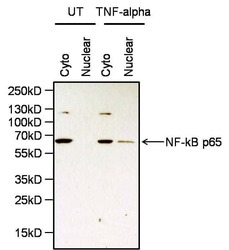
- Western blot analysis of NF-kB p65 was performed by loading 20 µg of cytoplasmic (cyto) and nuclear extracts from HeLa cells either left untreated (UT) or treated with 25 ng/mL of recombinant TNF-alpha (Product # RTNFAI) for 35 minutes at 37C, and 10 µL of PageRuler Plus Prestained Protein Ladder (Product # 26619) per well onto a 4-20% Tris-HCl polyacrylamide gel. Proteins were transferred to a PVDF membrane (Product # 88518) using the G2 Fast Blotter (Product # 62288), and blocked with StartingBlock T20 (TBS) Blocking Buffer (Product # 37543) for at least 1 hour at room temperature. NF-kB p65 was detected at 65 kD using an NF-kB p65 polyclonal antibody (Product # PA1-186) at a dilution of 1:2000 in StartingBlock T20 (TBS) Blocking Buffer overnight at 4C on a rocking platform, followed by an HRP-conjugated goat anti-rabbit IgG secondary antibody (Product # 31460) at a dilution of 1:40,000 for 30 minutes at room temperature. Chemiluminescent detection was performed using SuperSignal West Pico (Product # 34080).

- Submitted by
- Invitrogen Antibodies (provider)
- Main image
- Experimental details
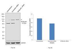
- Knockdown of NFkB p65 was achieved by transfecting HeLa with NFkB p65 specific siRNAs (Silencer® select Product # s11916, s11914). Western blot analysis was performed using Nuclear enriched extracts from the NFkB p65 knockdown cells (lane 3), scrambled siRNA transfected cells (lane 2), and untransfected cells (lane 1). The blot was probed with NFkB p65 Polyclonal Antibody (Product # PA1-186, 1 µg/mL) and Goat anti-Rabbit IgG (H+L) Superclonal™ Recombinant Secondary Antibody, HRP (Product # A27036, 1:20000 dilution). Decrease in signal upon siRNA mediated knock down confirms that antibody is specific to NFkB p65.

- Submitted by
- Invitrogen Antibodies (provider)
- Main image

- Experimental details
- Western blot analysis was performed on whole cell extracts (20 µg lysate) of NIH/3T3 (Lane 1), MCF7 (Lane 2), Hep G2 (Lane 3), Jurkat (lane 4) and HeLa (lane 5). The blots were probed with Anti-NF-kB p65 Rabbit Polyclonal Antibody (Product # PA1-186, 1-3 µg/mL) and detected by chemiluminescence using Goat anti-Rabbit IgG (H+L) Superclonal™ Secondary Antibody, HRP conjugate (Product # A27036, 0.4 µg/mL, 1:2500 dilution). A 60 kDa band corresponding to NF-kB p65 was observed across cell lines tested. Known quantity of protein samples were electrophoresed using Novex® NuPAGE® 4-12 % Bis-Tris gel (Product # NP0321BOX), XCell SureLock™ Electrophoresis System (Product # EI0002) and Novex® Sharp Pre-Stained Protein Standard (Product # LC5800). Resolved proteins were then transferred onto a nitrocellulose membrane with iBlot® 2 Dry Blotting System (Product # IB21001). The membrane was probed with the relevant primary and secondary Antibody following blocking with 5 % skimmed milk. Chemiluminescent detection was performed using Pierce™ ECL Western Blotting Substrate (Product # 32106).

Supportive validation
- Submitted by
- Invitrogen Antibodies (provider)
- Main image

- Experimental details
- Flow cytometry analysis of NF-kappa B p65 was done on HeLa cells. Cells were fixed with 70% ethanol for 10 minutes, permeabilized with 0.25% Triton™ X-100 for 20 minutes, and blocked with 5% BSA for 30 minutes at room temperature. Cells were labeled with NF-kappa B p65 Rabbit Polyclonal Antibody (PA1186, red histogram) or with rabbit isotype control (pink histogram) at 3-5 ug/million cells in 2.5% BSA. After incubation at room temperature for 2 hours, the cells were labeled with Alexa Fluor® 488 Goat Anti-Rabbit Secondary Antibody (A11008) at a dilution of 1:400 for 30 minutes at room temperature. The representative 10,000 cells were acquired and analyzed for each sample using an Attune® Acoustic Focusing Cytometer. The purple histogram represents unstained control cells and the green histogram represents no-primary-antibody control.

Supportive validation
- Submitted by
- Invitrogen Antibodies (provider)
- Main image

- Experimental details
- Enrichment of endogenous NF-kappa B p65 protein at specific gene loci using Anti-NF-kappa B p65 Rabbit Polyclonal Antibody: Chromatin Immunoprecipitation (ChIP) was performed using Anti-NF-kappa B p65 Rabbit Polyclonal Antibody (Product # PA1-186, 2 µg) on sheared chromatin from 2 million HeLa cells treated with 50 ng/mL of TNFalpha for 45 minutes using the "MAGnify ChIP system" kit (Product # 49-2024). Normal Rabbit IgG was used as a negative IP control. The purified DNA was analyzed by 7500 Fast qPCR system (Product # 4351106) with optimized PCR primer pairs for the promoter of active IL-8, IkB gene, used as positive control target, and the SAT2, used as negative control target. Data is presented as fold enrichment of the antibody signal versus the negative control IgG using the comparative CT method.

Supportive validation
- Submitted by
- Invitrogen Antibodies (provider)
- Main image

- Experimental details
- Immunoprecipitation of NF-kB p65 was performed on HeLa cells. Antigen-antibody complexes were formed by incubating 250 µg of HeLa whole cell lysate with 2.5 µg of an NF-kB p65 polyclonal antibody (Product # PA1-186) overnight on an end-over-end mixer at 4C. The immune complexes were captured on 100 µL Protein A/G Agarose (Product # 20421), washed extensively, and eluted with 5X Lane Marker Reducing Sample Buffer (Product # 39000). Samples were resolved on a 4-20% Tris-HCl polyacrylamide gel, transferred to a PVDF membrane (Product # 88518) using the G2 Fast Blotter (Product # 62288), and blocked with StartingBlock T20 (TBS) Blocking Buffer (Product # 37543) for at least 1 hour at room temperature. The membrane was probed with an NF-kB p65 polyclonal antibody (Product # PA1-186) at a dilution of 1:2000 overnight at 4C, washed in TBST, and probed with an HRP-conjugated mouse anti-rabbit light chain secondary antibody at a dilution of 1:40,000 for at least 30 minutes. Chemiluminescent detection was performed using SuperSignal West Dura (Product # 34075).

- Submitted by
- Invitrogen Antibodies (provider)
- Main image

- Experimental details
- Co-Immunoprecipitation of NF-kB p50 was performed on HeLa cells. Antigen-antibody complexes were formed by incubating 250 µg of HeLa whole cell lysate with 2.5 µg of an NF-kB p65 polyclonal antibody (Product # PA1-186) overnight on an end-over-end mixer at 4C. The immune complexes were captured on 100 µL Protein A/G Agarose (Product # 20421), washed extensively, and eluted with 5X Lane Marker Reducing Sample Buffer (Product # 39000). Samples were resolved on a 4-20% Tris-HCl polyacrylamide gel, transferred to a PVDF membrane (Product # 88518) using the G2 Fast Blotter (Product # 62288), and blocked with StartingBlock T20 (TBS) Blocking Buffer (Product # 37543) for at least 1 hour at room temperature. The membrane was probed with an NF-kB p50 rabbit monoclonal antibody at a dilution of 1:1000 overnight at 4C, washed in TBST, and probed with an HRP-conjugated mouse anti-rabbit light chain secondary antibody at a dilution of 1:40,000 for at least 30 minutes. Chemiluminescent detection was performed using SuperSignal West Dura (Product # 34075).

- Submitted by
- Invitrogen Antibodies (provider)
- Main image

- Experimental details
- NF-kB p65 was immunoprecipitated using 3 æg of the NF-kB p65 Rabbit Polyclonal Antibody (Product # PA1-186) from lysate of HeLa (Lane 3) using the Dynabeads® Protein A Immunoprecipitation Kit (Product # 10006D). Normal Rabbit IgG was used as a Isotype control (Lane 2). 10 % input represents the cell extract used for immunoprecipitation (Lane 1). Western blot analysis was performed using NF-kB p65 Rabbit Polyclonal Antibody (Product # PA1-186) and Goat anti-Rabbit IgG (H+L) Superclonal™ Secondary Antibody, HRP conjugate (Product # A27036, 0.4 æg/mL, 1:2500 dilution). Chemiluminescent detection was performed using Pierce™ ECL Western Blotting Substrate (Product # 32106).

- Submitted by
- Invitrogen Antibodies (provider)
- Main image

- Experimental details
- NULL

- Submitted by
- Invitrogen Antibodies (provider)
- Main image

- Experimental details
- NULL

- Submitted by
- Invitrogen Antibodies (provider)
- Main image

- Experimental details
- Fig. 5. FSTL1 overexpression rescues the miR-27a-mediated suppressive effect on RA-FLS migration and invasion by activating the TLR4/NFkappaB pathway. RA-FLS were transfected with miR-27a, exogenous FSTL1, or miR-27a and exogenous FSTL1. (A) The cell migration and invasion of RA-FLS as measured using the Transwell system. (B) Protein expression levels of TLR4 and NFkappaB in RA-FLS were detected by western blot analysis. * p < 0.05, versus control mimic group. # p < 0.05, versus Ad-beta-gal group. a p < 0.05, versus miR-27a mimic group.

- Submitted by
- Invitrogen Antibodies (provider)
- Main image

- Experimental details
- Figure 6 Melatonin attenuates NF-kBp65 upregulation by CoCl 2 in microglia. Western blot analysis ( a ) of NF-kBp65 levels in BV2 cells exposed to CoCl 2 (250 muM for 8 h) alone, or in combination with either melatonin (1 muM) or melatonin + EX527 (5 muM). In ( b ), immunostaining of NF-kBp65 (green) and nuclear counterstaining with DAPI (blue) following exposure to CoCl 2 alone (250 muM for 8 h), or in the presence of melatonin (1 muM) + EX527 (5 muM). Bars are mean +- SEM of three independent experiments. * p < 0.05 vs. control (C), deg p < 0.05 vs. CoCl 2 and # p < 0.05 vs. CoCl 2 + MEL by one-way ANOVA followed by Newman-Keuls test for statistical significance. Representative images are shown. Scale bar = 20 mum.

- Submitted by
- Invitrogen Antibodies (provider)
- Main image

- Experimental details
- Figure 10 Melatonin antagonises nuclear NF-kB localization in amoeboid microglia of the corpus callosum in rats subjected to CCAO. Seven-day-old rats were subjected to sham surgery or to ligation of the right carotid artery followed by hypoxia (HI) alone or with subsequent injection of melatonin (10 mg/kg; HI + melatonin (MEL). Animals were sacrificed after 24 h. Double immunohistochemical staining of Iba1 (red) and NF-kB (green) with DAPI nuclear counterstaining (blue) is shown. The graph reports the percentage of nuclear SIRT1+, Iba1+ microglial cells in the area. Representative images of ipsilateral CC are shown. Arrows indicate representative Iba+ cells with nuclear NF-kB; arrowheads indicate representative Iba1+ cells with extranuclear NF-kB staining. Asterisks indicate cells reported in the insets at higher magnification. Scale bar = 40 mum. Bars are mean +- SEM of at least three animals/group. * p < 0.05 vs. sham and ** p < 0.05 vs. HI by one-way ANOVA followed by Newman-Keuls test for statistical significance.

- Submitted by
- Invitrogen Antibodies (provider)
- Main image

- Experimental details
- Figure 4 Confocal microscopy for nuclear translocation of p65. (A) T47D cells (B) , MDA-MB-231 cells were pretreated with MyD88 inhibitor for 4 h prior to addition of TLR3 ligand (10 mug/ml) for 60 min. Cells were stained with antibody against p65 subunit of NF-kappaB and Alexa 594-tagged secondary antibody and counterstained with DAPI, and image acquired through confocal microscope (magnification, 63X). (C,D) Bar graph is presented as mean +- S.D for the quantitative measurements of nuclear localization of NF-kappaB at 30, 60, and 90 min of stimulation, analyzed through Image J package ( p < 0.05 is treated as significant). (E,F) Bar graph at 60 min of stimulation showing the highest nuclear localization of NF-kappaB.
