 Explore
 Validate
 Learn
Learn Western blot
 Immunoprecipitation
Antibody data
- Antibody Data
- Antigen structure
- References [16]
- Comments [0]
- Validations
- Western blot [4]
- Immunocytochemistry [1]
- Flow cytometry [1]
- Other assay [7]
Submit
Validation data
Reference
Comment
Report error
- Product number
- MA5-15160 - Provider product page

- Provider
- Invitrogen Antibodies
- Product name
- Phospho-NFkB p65 (Ser536) Monoclonal Antibody (T.849.2)
- Antibody type
- Monoclonal
- Antigen
- Synthetic peptide
- Description
- It is not recommended to aliquot this antibody. This antibody is not cross-reactive with the p50 subunit or other related proteins.
- Reactivity
- Human, Mouse, Rat, Hamster, Porcine
- Host
- Rabbit
- Isotype
- IgG
- Antibody clone number
- T.849.2
- Vial size
- 100 µL
- Concentration
- 57 µg/mL
- Storage
- -20°C
Submitted references Malaria parasite heme biosynthesis promotes and griseofulvin protects against cerebral malaria in mice.
B-cell antigen receptor expression and phosphatidylinositol 3-kinase signaling regulate genesis and maintenance of mouse chronic lymphocytic leukemia.
Capsaicin Attenuates Lipopolysaccharide-Induced Inflammation and Barrier Dysfunction in Intestinal Porcine Epithelial Cell Line-J2.
Fetuin-A secretion from β-cells leads to accumulation of macrophages in islets, aggravates inflammation and impairs insulin secretion.
βA1-crystallin regulates glucose metabolism and mitochondrial function in mouse retinal astrocytes by modulating PTP1B activity.
ECRG4 Represses Cell Proliferation and Invasiveness via NFIC/OGN/NF-κB Signaling Pathway in Bladder Cancer.
Hsp70 and NF-kB Mediated Control of Innate Inflammatory Responses in a Canine Macrophage Cell Line.
Interferon-γ signaling synergizes with LRRK2 in neurons and microglia derived from human induced pluripotent stem cells.
Roxatidine inhibits fibrosis by inhibiting NF‑κB and MAPK signaling in macrophages sensing breast implant surface materials.
IL4 Primes the Dynamics of Breast Cancer Progression via DUSP4 Inhibition.
An autocrine inflammatory forward-feedback loop after chemotherapy withdrawal facilitates the repopulation of drug-resistant breast cancer cells.
Twelve hours of heat stress induces inflammatory signaling in porcine skeletal muscle.
Cardamonin reduces chemotherapy-enriched breast cancer stem-like cells in vitro and in vivo.
β-Catenin and NF-κB co-activation triggered by TLR3 stimulation facilitates stem cell-like phenotypes in breast cancer.
Therapeutic potential of cannabidiol against ischemia/reperfusion liver injury in rats.
Coenzyme Q10 counteracts testicular injury induced by sodium arsenite in rats.
Chandana M, Anand A, Ghosh S, Das R, Beura S, Jena S, Suryawanshi AR, Padmanaban G, Nagaraj VA
Nature communications 2022 Jul 12;13(1):4028
Nature communications 2022 Jul 12;13(1):4028
B-cell antigen receptor expression and phosphatidylinositol 3-kinase signaling regulate genesis and maintenance of mouse chronic lymphocytic leukemia.
Schmid VK, Khadour A, Ahmed N, Brandl C, Nitschke L, Rajewsky K, Jumaa H, Hobeika E
Haematologica 2022 Aug 1;107(8):1796-1814
Haematologica 2022 Aug 1;107(8):1796-1814
Capsaicin Attenuates Lipopolysaccharide-Induced Inflammation and Barrier Dysfunction in Intestinal Porcine Epithelial Cell Line-J2.
Zhao X, Dong B, Friesen M, Liu S, Zhu C, Yang C
Frontiers in physiology 2021;12:715469
Frontiers in physiology 2021;12:715469
Fetuin-A secretion from β-cells leads to accumulation of macrophages in islets, aggravates inflammation and impairs insulin secretion.
Mukhuty A, Fouzder C, Kundu R
Journal of cell science 2021 Nov 1;134(21)
Journal of cell science 2021 Nov 1;134(21)
βA1-crystallin regulates glucose metabolism and mitochondrial function in mouse retinal astrocytes by modulating PTP1B activity.
Ghosh S, Liu H, Yazdankhah M, Stepicheva N, Shang P, Vaidya T, Hose S, Gupta U, Calderon MJ, Hu MW, Nair AP, Weiss J, Fitting CS, Bhutto IA, Gadde SGK, Naik NK, Jaydev C, Lutty GA, Handa JT, Jayagopal A, Qian J, Sahel JA, Rajasundaram D, Sergeev Y, Zigler JS Jr, Sethu S, Watkins S, Ghosh A, Sinha D
Communications biology 2021 Feb 24;4(1):248
Communications biology 2021 Feb 24;4(1):248
ECRG4 Represses Cell Proliferation and Invasiveness via NFIC/OGN/NF-κB Signaling Pathway in Bladder Cancer.
Liang X, Gao J, Wang Q, Hou S, Wu C
Frontiers in genetics 2020;11:846
Frontiers in genetics 2020;11:846
Hsp70 and NF-kB Mediated Control of Innate Inflammatory Responses in a Canine Macrophage Cell Line.
Lyu Q, Wawrzyniuk M, Rutten VPMG, van Eden W, Sijts AJAM, Broere F
International journal of molecular sciences 2020 Sep 4;21(18)
International journal of molecular sciences 2020 Sep 4;21(18)
Interferon-γ signaling synergizes with LRRK2 in neurons and microglia derived from human induced pluripotent stem cells.
Panagiotakopoulou V, Ivanyuk D, De Cicco S, Haq W, Arsić A, Yu C, Messelodi D, Oldrati M, Schöndorf DC, Perez MJ, Cassatella RP, Jakobi M, Schneiderhan-Marra N, Gasser T, Nikić-Spiegel I, Deleidi M
Nature communications 2020 Oct 14;11(1):5163
Nature communications 2020 Oct 14;11(1):5163
Roxatidine inhibits fibrosis by inhibiting NF‑κB and MAPK signaling in macrophages sensing breast implant surface materials.
Ji L, Wang T, Tian L, Song H, Gao M
Molecular medicine reports 2020 Jan;21(1):161-172
Molecular medicine reports 2020 Jan;21(1):161-172
IL4 Primes the Dynamics of Breast Cancer Progression via DUSP4 Inhibition.
Gaggianesi M, Turdo A, Chinnici A, Lipari E, Apuzzo T, Benfante A, Sperduti I, Di Franco S, Meraviglia S, Lo Presti E, Dieli F, Caputo V, Militello G, Vieni S, Stassi G, Todaro M
Cancer research 2017 Jun 15;77(12):3268-3279
Cancer research 2017 Jun 15;77(12):3268-3279
An autocrine inflammatory forward-feedback loop after chemotherapy withdrawal facilitates the repopulation of drug-resistant breast cancer cells.
Jia D, Li L, Andrew S, Allan D, Li X, Lee J, Ji G, Yao Z, Gadde S, Figeys D, Wang L
Cell death & disease 2017 Jul 13;8(7):e2932
Cell death & disease 2017 Jul 13;8(7):e2932
Twelve hours of heat stress induces inflammatory signaling in porcine skeletal muscle.
Ganesan S, Reynolds C, Hollinger K, Pearce SC, Gabler NK, Baumgard LH, Rhoads RP, Selsby JT
American journal of physiology. Regulatory, integrative and comparative physiology 2016 Jun 1;310(11):R1288-96
American journal of physiology. Regulatory, integrative and comparative physiology 2016 Jun 1;310(11):R1288-96
Cardamonin reduces chemotherapy-enriched breast cancer stem-like cells in vitro and in vivo.
Jia D, Tan Y, Liu H, Ooi S, Li L, Wright K, Bennett S, Addison CL, Wang L
Oncotarget 2016 Jan 5;7(1):771-85
Oncotarget 2016 Jan 5;7(1):771-85
β-Catenin and NF-κB co-activation triggered by TLR3 stimulation facilitates stem cell-like phenotypes in breast cancer.
Jia D, Yang W, Li L, Liu H, Tan Y, Ooi S, Chi L, Filion LG, Figeys D, Wang L
Cell death and differentiation 2015 Feb;22(2):298-310
Cell death and differentiation 2015 Feb;22(2):298-310
Therapeutic potential of cannabidiol against ischemia/reperfusion liver injury in rats.
Fouad AA, Jresat I
European journal of pharmacology 2011 Nov 16;670(1):216-23
European journal of pharmacology 2011 Nov 16;670(1):216-23
Coenzyme Q10 counteracts testicular injury induced by sodium arsenite in rats.
Fouad AA, Al-Sultan AI, Yacoubi MT
European journal of pharmacology 2011 Mar 25;655(1-3):91-8
European journal of pharmacology 2011 Mar 25;655(1-3):91-8
No comments: Submit comment
Supportive validation
- Submitted by
- Invitrogen Antibodies (provider)
- Main image
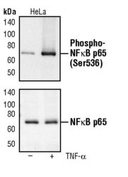
- Experimental details
- Western blot analysis of Phospho-NF-kappaB p65 pSer536 in extracts from HeLa cells, untreated or TNF-alpha treated (20 ng/mL for 5 minutes), using Phospho-NF-kappaB p65 pSer536 monoclonal antibody (Product # MA5-15160) (upper) or a NF-kappa-B p65 polyclonal antibody (lower).

- Submitted by
- Invitrogen Antibodies (provider)
- Main image
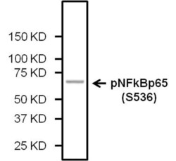
- Experimental details
- J-Lat Full Length cell lysate 40 µg was loaded on 4-15% gel and transferred to 0.45 µm PVDF Membrane. Phospho-NFkB p65 (Ser536) Monoclonal Antibody (Product # MA5-15160) was diluted at 1:500 in 1xTBST buffer with 5% BSA and incubated overnight at 4C. Goat anti Rabbit HRP-conjugated secondary antibody was diluted at 1:2000 in 1xTBST buffer with 5% non fat milk and incubated for 1 hour at 21C.

- Submitted by
- Invitrogen Antibodies (provider)
- Main image
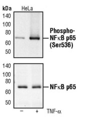
- Experimental details
- Western blot analysis of Phospho-NF-kappaB p65 pSer536 in extracts from HeLa cells, untreated or TNF-alpha treated (20 ng/mL for 5 minutes), using Phospho-NF-kappaB p65 pSer536 monoclonal antibody (Product # MA5-15160) (upper) or a NF-kappa-B p65 polyclonal antibody (lower).

- Submitted by
- Invitrogen Antibodies (provider)
- Main image
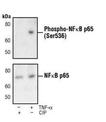
- Experimental details
- Western blot analysis of Phospho-NFkB p65 (Ser536) using a monoclonal antibody (Product # MA5-15160).

Supportive validation
- Submitted by
- Invitrogen Antibodies (provider)
- Main image

- Experimental details
- Immunofluorescent analysis of Phospho-NF-kappaB p65 pSer536 in HeLa cells, TNF-alpha treated (20 ng/mL for 20 min), using a Phospho-NF-kappaB p65 pSer536 monoclonal antibody (Product # MA5-15160) (green). Actin filaments are labeled with a fluorescent red phalloidin.

Supportive validation
- Submitted by
- Invitrogen Antibodies (provider)
- Main image

- Experimental details
- Flow cytometric analysis of Phospho-NF-kappaB p65 pSer536 in untreated (blue) or TNF-alpha-treated (green, 20 ng/mL for 10 minutes) HeLa cells using a Phospho-NF-kappaB p65 pSer536 monoclonal antibody (Product # MA5-15160) compared to a nonspecific negative control antibody (red).

Supportive validation
- Submitted by
- Invitrogen Antibodies (provider)
- Main image

- Experimental details
- Figure 4 NF- kappa B signal is not exclusively responsible for enhanced CSC phenotypes after TLR3 activation. ( a ) SUM190, SUM149, BT483, and Cama-1 cells were treated with 1 mu g/ml poly(I:C) for 4 days. Nuclear extracts (Nu) and cytoplasmic extracts (Cyto) were immunoblotted to examine nuclear translocation of NF- kappa B p65. alpha -tubulin and Lamin A/C: internal loading controls for cytoplasmic and nuclear proteins, respectively. ( b ) qPCR analysis of target genes of NF- kappa B pathway: IL8 and I kappa B alpha in SUM190, SUM149, BT483, and Cama-1 following 1 mu g/ml of poly(I:C) treatment for 4 days. Data represent the average+-S.D., n =3; * P

- Submitted by
- Invitrogen Antibodies (provider)
- Main image

- Experimental details
- Figure 6 Cardamonin blocks both beta -catenin and NF- kappa B pathways and abrogates TLR3-activation-induced CSCs in vitro . ( a ) Molecular structure of cardamonin (2,4-dihydroxy-6-methoxychalcone) ( b ) Representative images of SUM190 cells treated with 1 mu g/ml of poly(I:C) in the presence or absence of cardamonin (10 mu M) for 4 days. Cardamonin abrogates the formation of non-adherent spherical clusters induced by poly(I:C) stimulation. Scale bars, 100 mu m. ( c ) Western blot analysis of nuclear translocation of NF- kappa B p65 and active beta -catenin. SUM190 cells were pretreated with 10 mu M of cardamonin for 4 h, followed by stimulation with poly(I:C) (1 mu g/ml) for 4 days in the presence of cardamonin. alpha -tubulin and Lamin A/C: internal loading controls for cytoplasmic (Cyto) and nuclear (Nu) proteins, respectively. ( d ) Flow-cytometry analysis of CD44 high /CD24 -/low sub-population in four different breast cancer cell lines treated as described in ( c ). Data represent the average+-S.D., n =3; * P

- Submitted by
- Invitrogen Antibodies (provider)
- Main image

- Experimental details
- Figure 5 beta -Catenin and NF-kappaB co-mediate TLR3 activation-induced CSC phenotypes. ( a ) qPCR analysis of target genes of beta -catenin pathway (Axin2 and CylinD1) in SUM190 after treatment with 1 mu g/ml of poly(I:C) for 4 days. Data represent the average+-S.D., n =5; * P

- Submitted by
- Invitrogen Antibodies (provider)
- Main image

- Experimental details
- Figure 4 The effect of cell stress on NF-kappaB phosphorylation in LPS-stimulated 030D cells. The 030D cells were incubated with different concentrations (1.25 and 2.5 uM) of arsenite, or without, for 16 h, after which the cells were exposed to LPS and harvested at 5 min ( A , E ), 15 min ( B , F ), 30 min ( C , G ) and 60 min ( D , H ). Whole cell lysates were extracted and analyzed by Western Blotting. Control: untreated cells. Phosphorylated NF-kappaB and total NF-kappaB were detected with rabbit monoclonal anti-phospho-NF-kappaB p65 and HRP-labeled swine-anti rabbit IgG, and mouse monoclonal anti- NF-kappaB p65 and HRP-labeled rabbit anti-mouse IgG, respectively. The densitometry of the protein bands was scanned and quantitated with Image lab TM software 6.0.1 ( E - H ). The total NF-kappaB levels were used as an internal control. Data are shown as the mean +- SD and are representative of three independent experiments. * p < 0.05, ** p < 0.01, and *** p < 0.001, vs. LPS alone group.

- Submitted by
- Invitrogen Antibodies (provider)
- Main image

- Experimental details
- Figure 6 The effect of a deficiency of inducible Hsp70 on pro-inflammatory cytokine expression and NF-kappaB phosphorylation. Hsp70 knockout 030D cells were treated with different concentrations (1.25, 2.5 or 5 uM) of arsenite, or without, for 16 h, and then exposed to LPS. The cells were harvested after 6 h ( A - C ) of LPS exposure. qPCR was performed to detect the expression of IL-6 ( A ), IL-1beta ( B ) and TNF-alpha ( C ). For Western Blotting, the cells were harvested after 5 ( D , H ), 15 ( E , I ), 30 ( F , J ) and 60 ( G , K ) min of LPS exposure. Western Blotting was performed to detect levels of phosphorylated NF-kappaB and total NF-kappaB at certain time points. The densitometry of the protein bands was scanned and quantitated with Image lab TM software 6.0.1. ( H - K ). The total NF-kappaB levels were used as an internal control. Data are shown as the mean +- SD and are representative of three independent experiments. * p < 0.05, ** p < 0.01 vs. LPS alone group.

- Submitted by
- Invitrogen Antibodies (provider)
- Main image

- Experimental details
- Figure 4 Effects of capsaicin and LPS treatment on the phosphorylation of NF-kappaB p65 in LPS-challenged IPEC-J2 cells. The level of phosphorylated NF-kappaB p65 was evaluated using Immunofluorescence staining (A) and Western blotting (B) , respectively. For Immunofluorescence staining, IPEC-J2 cells were seeded into coverslips at a density of 1x10 5 /well and cultured for 2 weeks. After being pretreated with capsaicin (100 muM), cells were stimulated with LPS. Cells then were then fixed for phosphorylated NF-kappaB p65 staining as described in the Materials and Methods. As for Western blot, IPEC-J2 cells were cultured in 6-well plates. Cells were firstly pre-treated with capsaicin (100 muM) for 2 h and then stimulated with LPS (10 mug/mL) for 6 h. The protein was extracted and the total NF-kappaB p65, as well as phosphorylation of NF-kappaB p65 in LPS-challenged IPEC-J2 cells were detected as described in the Materials and Methods. Values are presented as mean +- SEM. Different letters on bars (a, b) indicate significant differences, P < 0.05.

- Submitted by
- Invitrogen Antibodies (provider)
- Main image
- Experimental details
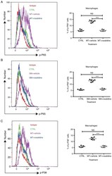
- Figure 5. Addition of roxatidine to the culture medium of RAW 264.7 macrophages inhibited the activation of the NF-kappaB and MAPK signaling pathways. RAW 264.7 macrophages were cultured in serum-free media at 37degC. Then, 24 h after cells were seeded, roxatidine (25 uM) was added to the media 1 h prior to stimulation with silicone surface materials. Cells were collected for analysis of phosphorylation of NF-kappaB subunit p65 and p38 MAPK by flow cytometry 15 minutes after adding silicone surface materials to culture media. Administration of roxatidine suppressed p65 activation stimulated by (A) MT and (B) SM, as well as p38 phosphorylation stimulated by (C) MT. p38 phosphorylation stimulated by (D) SM. n=6 wells/group, from one of triplicated experiments. n.s., not significant and ***P
