 Explore
 Validate
 Learn
Learn Western blot
 ELISA
ELISAAntibody data
- Antibody Data
- Antigen structure
- References [2]
- Comments [0]
- Validations
- Western blot [2]
- Immunohistochemistry [1]
Submit
Validation data
Reference
Comment
Report error
- Product number
- NBP1-77815 - Provider product page

- Provider
- Novus Biologicals
- Proper citation
- Novus Cat#NBP1-77815, RRID:AB_11016281
- Product name
- Mouse Monoclonal RelA/NFkB p65 Antibody
- Antibody type
- Monoclonal
- Description
- Protein A purified.
- Reactivity
- Human
- Host
- Mouse
- Isotype
- IgG
- Vial size
- 0.1 mg
- Concentration
- 1.0 mg/ml
- Storage
- Aliquot and store at -20C or -80C. Avoid freeze-thaw cycles.
Submitted references The flavonoid apigenin reduces prostate cancer CD44(+) stem cell survival and migration through PI3K/Akt/NF-κB signaling.
Her2 activates NF-kappaB and induces invasion through the canonical pathway involving IKKalpha.
Erdogan S, Doganlar O, Doganlar ZB, Serttas R, Turkekul K, Dibirdik I, Bilir A
Life sciences 2016 Oct 1;162:77-86
Life sciences 2016 Oct 1;162:77-86
Her2 activates NF-kappaB and induces invasion through the canonical pathway involving IKKalpha.
Merkhofer EC, Cogswell P, Baldwin AS
Oncogene 2010 Feb 25;29(8):1238-48
Oncogene 2010 Feb 25;29(8):1238-48
No comments: Submit comment
Supportive validation
- Submitted by
- Novus Biologicals (provider)
- Main image
- Experimental details
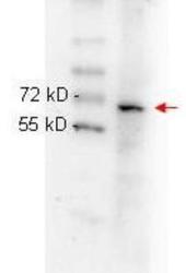
- Western Blot: RelA/NFkB p65 Antibody (27F9.G4) [NBP1-77815] - This antibody was used in HeLa whole cell lysate. Lysate was run on 4-20% gradient gel transferred under standard conditions and blocked in 1% BSA-TTBS 30 min RT. Blot was probed with monoclonal anti p65 1:1000 in 1% BSA-TBS-T o/n 4C and detected with HRP conjugated Rb-anti Mouse antibody 1:40,000 in MB-070 30 min RT.
- Submitted by
- Novus Biologicals (provider)
- Main image
- Experimental details
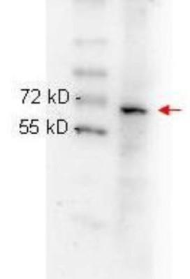
- Western Blot: RelA/NFkB p65 Antibody (27F9.G4) [NBP1-77815] - Samples were prepared in RIPA lysis buffer, boiled with NuPage 4x LDS Sample Buffer and run on NuPage 4-12% Bis-Tris Gels. Blot was incubated with primary antibody at a dilution of 1:500 and detected with HRP conjugated anti mouse antibody at a dilution of 1:10000. Image provided courtesy of Dr. Al Baldwin, University of North Carolina, Chapel Hill.

Supportive validation
- Submitted by
- Novus Biologicals (provider)
- Main image
- Experimental details
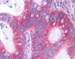
- Immunohistochemistry-Paraffin: RelA/NFkB p65 Antibody (27F9.G4) [NBP1-77815] - Human tissues. showed moderate to strong staining within many tissues, including epithelium of the breast, small intestine, kidney, pancreas, prostate, skin, placenta, and uterus, as well as within neurons and lymphoid tissues such as spleen, thymus, and tonsil. The antibody produced an excellent signal with almost no background staining at a concentration of 2.5 ug/ml. The image displayed shows specific staining in colon carcinoma as the precipitated red signal, with a hematoxylin purple nuclear counterstain.
