 Explore
 Validate
 Learn
Learn Western blot
Antibody data
- Antibody Data
- Antigen structure
- References [1]
- Comments [0]
- Validations
- Western blot [1]
- Immunocytochemistry [1]
Submit
Validation data
Reference
Comment
Report error
- Product number
- MAB7226 - Provider product page

- Provider
- R&D Systems
- Product name
- Human Phospho-RelA/NFkB p65 (S536) Antibody
- Antibody type
- Monoclonal
- Description
- Protein A or G purified from hybridoma culture supernatant. Detects human RelA/NF kappa B p65 when phosphorylated at S536.
- Reactivity
- Human
- Host
- Mouse
- Conjugate
- Unconjugated
- Isotype
- IgG
- Antibody clone number
- 817403
- Vial size
- 100 ug
- Concentration
- LYOPH
- Storage
- Use a manual defrost freezer and avoid repeated freeze-thaw cycles. 12 months from date of receipt, -20 to -70 °C as supplied. 1 month, 2 to 8 °C under sterile conditions after reconstitution. 6 months, -20 to -70 °C under sterile conditions after reconstitution.
Submitted references Protective effect of KLF15 on vascular endothelial dysfunction induced by TNF‑α.
Liu B, Xu L, Yu X, Li W, Sun X, Xiao S, Guo M, Wang H
Molecular medicine reports 2018 Aug;18(2):1987-1994
Molecular medicine reports 2018 Aug;18(2):1987-1994
No comments: Submit comment
Supportive validation
- Submitted by
- R&D Systems (provider)
- Main image

- Experimental details
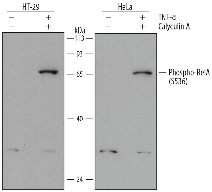
- Detection of Human Phospho-RelA/NF kappa B p65 (S536) by Western Blot. Western blot shows lysates of HT-29 human colon adenocarcinoma cell line and HeLa human cervical epithelial carcinoma cell line untreated (-) or treated (+) with 100 nM Calyculin A (Catalog # 1336) and 20 ng/mL Recombinant Human TNF-alpha (Catalog # 210-TA) for 10 minutes. PVDF membrane was probed with 2 µg/mL of Mouse Anti-Human Phospho-RelA/NF kappa B p65 (S536) Monoclonal Antibody (Catalog # MAB7226) followed by HRP-conjugated Anti-Mouse IgG Secondary Antibody (Catalog # HAF018). A specific band was detected for Phospho-RelA/ NF kappa B p65 (S536) at approximately 65 kDa (as indicated). This experiment was conducted under reducing conditions and using Immunoblot Buffer Group 2.
Supportive validation
- Submitted by
- R&D Systems (provider)
- Main image

- Experimental details
- Phospho-RelA/NFkB p65 (S536) in HT-29 Human Cell Line. RelA/NF kappa B p65 phosphorylated at S536 was detected in immersion fixed HT-29 human colon adenocarcinoma cell line untreated (lower panel) or treated with Calyculin A (Catalog # 1336) and Recombinant Human TNF-alpha (Catalog # 210-TA; upper panel) using Mouse Anti-Human Phospho-RelA/NF kappa B p65 (S536) Monoclonal Antibody (Catalog # MAB7226) at 2 µg/mL for 3 hours at room temperature. Cells were stained using the NorthernLights™ 557-conjugated Anti-Mouse IgG Secondary Antibody (red; Catalog # NL007) and counterstained with DAPI (blue). Specific staining was localized to cytoplasm and cell surfaces. View our protocol for Fluorescent ICC Staining of Cells on Coverslips.
