Explore
 Validate
 Learn
Learn Western blot
 Immunocytochemistry
Antibody data
- Antibody Data
- Antigen structure
- References [4]
- Comments [0]
- Validations
- Western blot [4]
- Immunocytochemistry [2]
- Immunoprecipitation [1]
- Immunohistochemistry [1]
- Chromatin Immunoprecipitation [1]
Submit
Validation data
Reference
Comment
Report error
- Product number
- GTX109679 - Provider product page

- Provider
- GeneTex
- Proper citation
- GeneTex Cat#GTX109679, RRID:AB_1950482
- Product name
- HDAC3 antibody [C3], C-term
- Antibody type
- Polyclonal
- Reactivity
- Human, Mouse, Rat
- Host
- Rabbit
Submitted references NKILA lncRNA promotes tumor immune evasion by sensitizing T cells to activation-induced cell death.
Inhibition of HDAC3- and HDAC6-Promoted Survivin Expression Plays an Important Role in SAHA-Induced Autophagy and Viability Reduction in Breast Cancer Cells.
Exercise increases the binding of MEF2A to the Cpt1b promoter in mouse skeletal muscle.
NudC deacetylation regulates mitotic progression.
Huang D, Chen J, Yang L, Ouyang Q, Li J, Lao L, Zhao J, Liu J, Lu Y, Xing Y, Chen F, Su F, Yao H, Liu Q, Su S, Song E
Nature immunology 2018 Oct;19(10):1112-1125
Nature immunology 2018 Oct;19(10):1112-1125
Inhibition of HDAC3- and HDAC6-Promoted Survivin Expression Plays an Important Role in SAHA-Induced Autophagy and Viability Reduction in Breast Cancer Cells.
Lee JY, Kuo CW, Tsai SL, Cheng SM, Chen SH, Chan HH, Lin CH, Lin KY, Li CF, Kanwar JR, Leung EY, Cheung CC, Huang WJ, Wang YC, Cheung CH
Frontiers in pharmacology 2016;7:81
Frontiers in pharmacology 2016;7:81
Exercise increases the binding of MEF2A to the Cpt1b promoter in mouse skeletal muscle.
Yuan H, Niu Y, Liu X, Fu L
Acta physiologica (Oxford, England) 2014 Dec;212(4):283-92
Acta physiologica (Oxford, England) 2014 Dec;212(4):283-92
NudC deacetylation regulates mitotic progression.
Chuang C, Pan J, Hawke DH, Lin SH, Yu-Lee LY
PloS one 2013;8(9):e73841
PloS one 2013;8(9):e73841
No comments: Submit comment
Enhanced validation
Supportive validation
- Submitted by
- GeneTex (provider)
- Enhanced method
- Genetic validation
- Main image
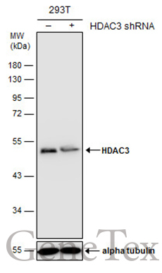
- Experimental details
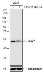
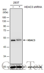
- Non-transfected (¡V) and transfected (+) 293T whole cell extracts (30 ?g) were separated by 10% SDS-PAGE, and the membrane was blotted with HDAC3 antibody [C3], C-term (GTX109679) diluted at 1:500. The HRP-conjugated anti-rabbit IgG antibody (GTX213110-01) was used to detect the primary antibody.
Supportive validation
- Submitted by
- GeneTex (provider)
- Main image
- Experimental details
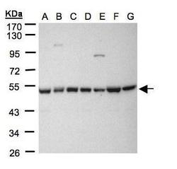
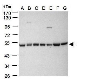
- Sample(30 ?g whole cell lysate)A: 293TB: A431 (GTX27909)C: H1299D: HeLa S3 (GTX14654)E: HepG2 (GTX27900)F: MOLT4 (GTX27912)G: Raji (GTX27908)10% SDS PAGEGTX109679 diluted at 1:1000The HRP-conjugated anti-rabbit IgG antibody (GTX213110-01) was used to detect the primary antibody.
- Submitted by
- GeneTex (provider)
- Main image
- Experimental details
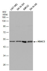
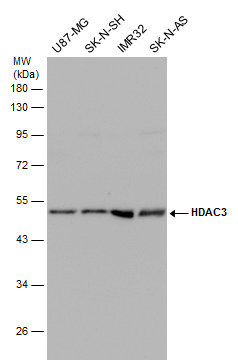
- Various whole cell extracts (30 ?g) were separated by 10% SDS-PAGE, and the membrane was blotted with HDAC3 antibody [C3], C-term (GTX109679) diluted at 1:500. The HRP-conjugated anti-rabbit IgG antibody (GTX213110-01) was used to detect the primary antibody.
- Submitted by
- GeneTex (provider)
- Main image
- Experimental details
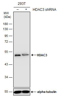
- Non-transfected (¡V) and transfected (+) 293T whole cell extracts (30 ?g) were separated by 10% SDS-PAGE, and the membrane was blotted with HDAC3 antibody [C3], C-term (GTX109679) diluted at 1:500. The HRP-conjugated anti-rabbit IgG antibody (GTX213110-01) was used to detect the primary antibody.
Supportive validation
- Submitted by
- GeneTex (provider)
- Main image
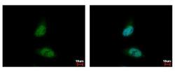
- Experimental details
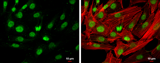
- HDAC3 antibody [C3], C-term detects HDAC3 protein at nucleus by immunofluorescent analysis. Sample: HeLa cells were fixed in 4% paraformaldehyde at RT for 15 min.Green: HDAC3 protein stained by HDAC3 antibody [C3], C-term (GTX109679) diluted at 1:500.Blue: Hoechst 33342 staining.
- Submitted by
- GeneTex (provider)
- Main image
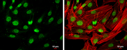
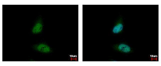
- Experimental details
- HDAC3 antibody [C3], C-term detects HDAC3 protein at nucleus by immunofluorescent analysis.Sample: SK-N-SH cells were fixed in 4% paraformaldehyde at RT for 15 min.Green: HDAC3 protein stained by HDAC3 antibody [C3], C-term (GTX109679) diluted at 1:400.Red: Phalloidin, a cytoskeleton marker, diluted at 1:200.Scale bar = 10 £gm.
Supportive validation
- Submitted by
- GeneTex (provider)
- Main image
- Experimental details
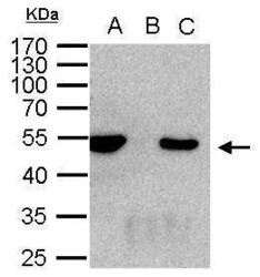
- HDAC3 antibody immunoprecipitates HDAC3 protein in IP experiments. IP Sample: 1000 ?g 293T whole cell lysate/extract A. 50 £gg 293T whole cell lysate/extract B. Control with 2 £gg of preimmune rabbit IgG C. Immunoprecipitation of HDAC3 protein by 2 £gg of HDAC3 antibody (GTX109679) 10% SDS-PAGE The immunoprecipitated HDAC3 protein was detected by HDAC3 antibody (GTX109679) diluted at 1:1000. EasyBlot anti-rabbit IgG (GTX221666-01) was used as a secondary reagent.

Supportive validation
- Submitted by
- GeneTex (provider)
- Main image
- Experimental details
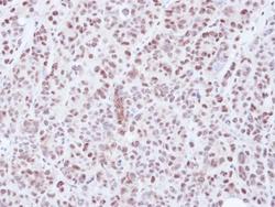
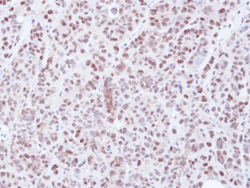
- Immunohistochemical analysis of paraffin-embedded SW480 xenograft, using HDAC3(GTX109679) antibody at 1:500 dilution.
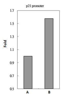
Supportive validation
- Submitted by
- GeneTex (provider)
- Main image
- Experimental details
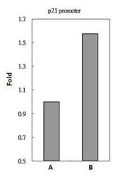
- HDAC3 antibody immunoprecipitates HDAC3 protein-DNA in ChIP experiments. ChIP Sample: 293T whole cell lysate/extract A. 5 £gg preimmune rabbit IgG B. 5 £gg of HDAC3 antibody (GTX109679) The precipitated DNA was detected by PCR with primer set targeting to p21 promoter.