 Explore
 Validate
 Learn
Learn Western blot
Antibody data
- Antibody Data
- Antigen structure
- References [2]
- Comments [0]
- Validations
- Western blot [6]
- Immunocytochemistry [3]
- Immunohistochemistry [1]
- Other assay [5]
Submit
Validation data
Reference
Comment
Report error
- Product number
- PA5-20007 - Provider product page

- Provider
- Invitrogen Antibodies
- Product name
- PUMA alpha Polyclonal Antibody
- Antibody type
- Polyclonal
- Antigen
- Synthetic peptide
- Description
- The PA5-20007 immunogen is located within the last 50 amino acids of PUMA. The immunogenic sequence is identical between the A and B forms of PUMA.
- Concentration
- 1 mg/mL
Submitted references Circular RNA circLDB2 functions as a competing endogenous RNA to suppress development and promote cisplatin sensitivity in non-squamous non-small cell lung cancer.
Aberrant Caspase Activation in Laminin-α2-Deficient Human Myogenic Cells is Mediated by p53 and Sirtuin Activity.
Wang Y, Li L, Zhang W, Zhang G
Thoracic cancer 2021 Jul;12(13):1959-1972
Thoracic cancer 2021 Jul;12(13):1959-1972
Aberrant Caspase Activation in Laminin-α2-Deficient Human Myogenic Cells is Mediated by p53 and Sirtuin Activity.
Yoon S, Beermann ML, Yu B, Shao D, Bachschmid M, Miller JB
Journal of neuromuscular diseases 2018;5(1):59-73
Journal of neuromuscular diseases 2018;5(1):59-73
No comments: Submit comment
Supportive validation
- Submitted by
- Invitrogen Antibodies (provider)
- Main image
- Experimental details
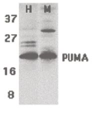
- Western blot analysis of human (H) K562 and mouse (M) 3T3 cell lysates using a PUMA polyclonal antibody (Product # PA5-20007) at 2 µg/mL

- Submitted by
- Invitrogen Antibodies (provider)
- Main image

- Experimental details
- Western Blot Validation of PUMA in K562 and 3T3/NIH Cells. Loading: 15 µg of lysates per lane. Antibodies: PUMA Polyclonal Antibody (Product # PA5-20007) (2 µg/mL), 1 h incubation at RT in 0.05 NFDM/TBST. Secondary: Goat anti-rabbit IgG HRP conjugate at 1:10,000 dilution.

- Submitted by
- Invitrogen Antibodies (provider)
- Main image

- Experimental details
- Western Blot analysis of PUMA in Tet cells using PUMA Polyclonal Antibody (Product # PA5-20007) (Han et al., 2010). Tet-induced p53 cells were treated with NOXA, Puma, Bim or non-targeting siRNAs that were utilized in this experiment. PUMA protein levels were markedly reduced in PUMA KD cells detected by anti-PUMA antibodies.

- Submitted by
- Invitrogen Antibodies (provider)
- Main image

- Experimental details
- Western Blot analysis of PUMA in 2983 Cells. Lysates/proteins (15 µg per lane) were loaded onto SDS-PAGE. The membrane was probed with PUMA Polyclonal Antibody (Product # PA5-20007) (lane 1-3: 1, 2 and 4 µg/mL). 1 h incubation at RT in 5% NFDM/TBST. Secondary: Goat anti-rabbit IgG HRP conjugate at 1:10,000 dilution.

- Submitted by
- Invitrogen Antibodies (provider)
- Main image

- Experimental details
- Western Bloat analysis PUMA siRNA Knockdown in 293 Cells. 293 cells were transfected with control siRNAs (lane 1) or PUMA siRNAs (lane 2) Loading: 15 µg of 293 whole cell lysates per lane. Antibodies: PUMA Polyclonal Antibody (Product # PA5-20007) (2 µg/mL), beta-actin (1 µg/mL) and GAPDH (0.02 µg/mL), 1 h incubation at RT in 5% NFDM/TBST. Secondary: Goat anti-rabbit IgG HRP conjugate at 1:10,000 dilution.

- Submitted by
- Invitrogen Antibodies (provider)
- Main image

- Experimental details
- Western Blot Validation of PUMA in K562 and 3T3/NIH Cells. Loading: 15 µg of lysates per lane. Antibodies: PUMA Polyclonal Antibody (Product # PA5-20007) (2 µg/mL), 1 h incubation at RT in 0.05 NFDM/TBST. Secondary: Goat anti-rabbit IgG HRP conjugate at 1:10,000 dilution.

Supportive validation
- Submitted by
- Invitrogen Antibodies (provider)
- Main image

- Experimental details
- Immunofluorescent analysis of K562 cells using a PUMA polyclonal antibody (Product # PA5-20007) at a 2 µg/mL dilution.

- Submitted by
- Invitrogen Antibodies (provider)
- Main image

- Experimental details
- Immunocytochemistry of K562 cells using PUMA Polyclonal Antibody (Product # PA5-20007) at 1 µg/mL. Cells were fixed with formaldehyde and blocked with 0.1 serum for 1 h at RT; antigen retrieval was by heat mediation with a citrate buffer (pH6). Samples were incubated with primary antibody overnight at 4°C. A goat anti-rabbit IgG H&L (HRP) at 1:250 was used as secondary. Counter stained with Hematoxylin.

- Submitted by
- Invitrogen Antibodies (provider)
- Main image

- Experimental details
- Immunofluorescent analysis of 4% paraformaldehyde-fixed K562 cells labeling PUMA with PUMA Polyclonal Antibody (Product # PA5-20007) at 2 µg/mL, followed by goat anti-rabbit IgG secondary antibody at 1:500 dilution (red). Image showing cytosol staining on K562 cells.

Supportive validation
- Submitted by
- Invitrogen Antibodies (provider)
- Main image

- Experimental details
- Immunocytochemistry staining of K562 cells using a PUMA polyclonal antibody (Product # PA5-20007) at a 1 µg/mL dilution.

Supportive validation
- Submitted by
- Invitrogen Antibodies (provider)
- Main image

- Experimental details
- NULL

- Submitted by
- Invitrogen Antibodies (provider)
- Main image

- Experimental details
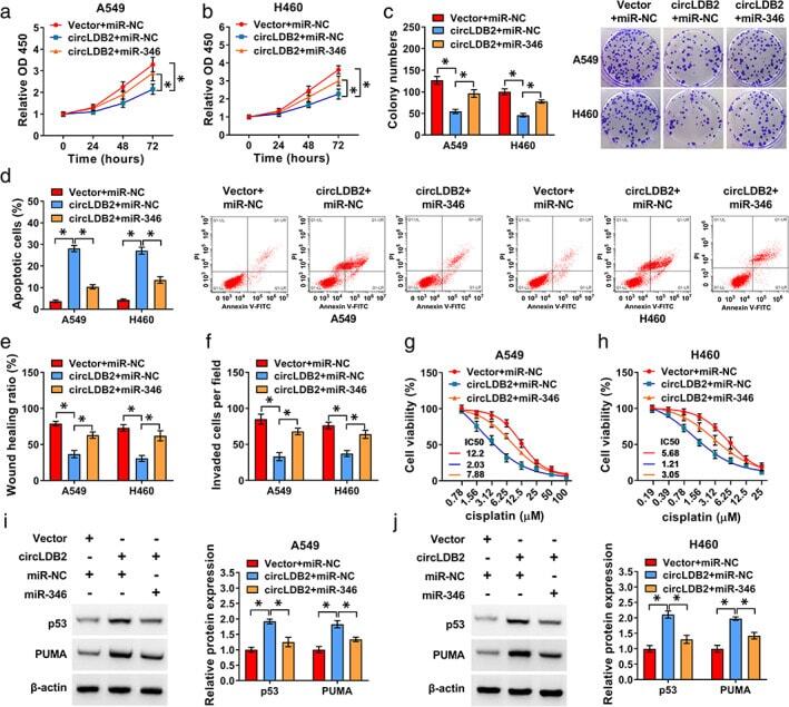
- FIGURE 5 CircLDB2 regulated non-squamous NSCLC development and cisplatin sensitivity in vitro by miR-346. Vector-transduced or circLDB2-infected A549 and H460 cells were transfected with miR-NC mimic or miR-346 mimic. (a) and (b) CCK-8 assay of cell proliferation. (c) Representative images depicting a colony formation assay. (d) Representative images showing a cell apoptosis assay and flow cytometry for cell apoptosis. (e) Wound-healing assay of cell migration. (f) Cell invasion by transwell assay. (g) and (h) transfected cells were exposed to various concentrations of cisplatin, followed by the evaluation of cell viability by CCK-8 assay. (i) and (j) Western blot showing the levels of p53 and PUMA in transfected cells. * p < 0.05
- Submitted by
- Invitrogen Antibodies (provider)
- Main image
- Experimental details
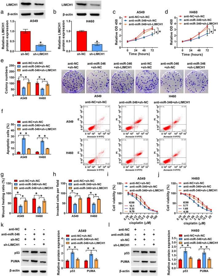
- FIGURE 7 LIMCH1 was a functional target of miR-346. (a) and (b) Western blot analysis of LIMCH1 protein level in cells transfected with sh-NC or sh-LIMCH1. A549 and H460 cells were transfected with anti-NC + sh-NC, anti-miR-346 + sh-NC or anti-miR-346 + sh-LIMCH1. (c) and (d) CCK-8 assay of proliferation of transfected cells. (e) Representative images depicting a colony formation assay. (f) Representative images depicting a cell apoptosis assay and cell apoptosis by flow cytometry. (g) Wound-healing assay of cell migration. (h) Transwell assay of cell invasion. (i) and (j) CCK-8 assay for viability of transfected cells under cisplatin exposure. (k) and (l) Western blot showing the levels of p53 and PUMA in transfected cells. * p < 0.05
- Submitted by
- Invitrogen Antibodies (provider)
- Main image
- Experimental details
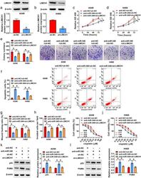
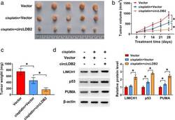
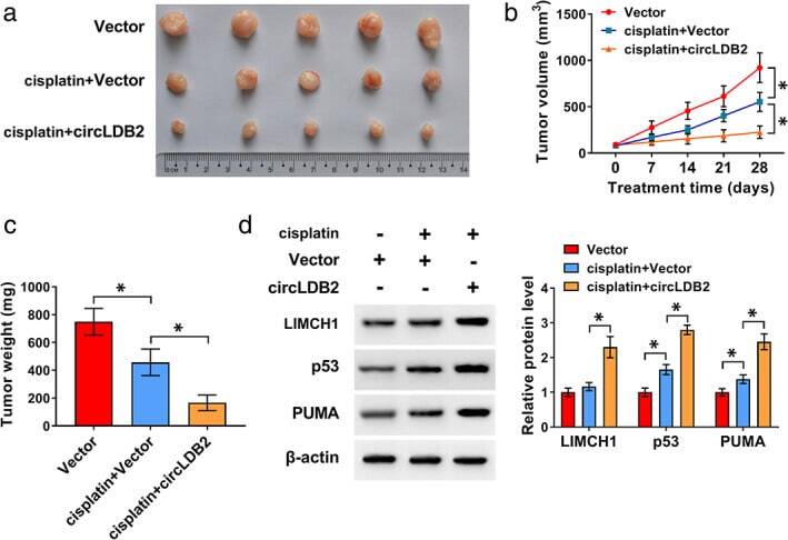
- FIGURE 8 Overexpression of circLDB2 promoted the antitumor effect of cisplatin in vivo. The xenograft tumors were generated by subcutaneous injection of vector-transduced or circLDB2-transduced A549 cells (2 x 10 6 per mouse, n = 5 per group), and cisplatin treatment was performed twice a week by intraperitoneal injection. Mice were terminated at day 28 after cell implantation. (a) Representative images of the xenograft tumors. (b) Growth curves of the excised tumors. (c) Tumor average weight of the xenograft tumors. (d) Western blot showing the levels of LIMCH1, p53, and PUMA in the excised tumors. * p < 0.05
- Submitted by
- Invitrogen Antibodies (provider)
- Main image

- Experimental details
- FIGURE 3 CircLDB2 overexpression regulated cell proliferation, apoptosis, migration, invasion, and cisplatin sensitivity in vitro. A549 and H460 cells were transduced with negative control lentivirus (vector) or circLDB2 overexpression lentivirus (circLDB2). (a) and (b) CCK-8 assay of proliferation of transduced cells. (c) Representative images depicting a colony formation assay and colony formation assay of colony formation ability. (d) Representative images depicting a cell apoptosis assay and cell apoptosis by flow cytometry. (e) Representative images showing a cell migration assay and cell migration by wound-healing assay. (f) Representative images showing a cell invasion assay and cell invasion by transwell assay. (g) and (h) CCK-8 assay of viability of transduced cells under cisplatin (various doses) exposure. (i) and (j) representative images depicting a western blot assay and western blot showing the levels of p53 and PUMA in transduced cells. * p < 0.05
