 Explore
 Validate
 Learn
Learn Western blot
Antibody data
- Antibody Data
- Antigen structure
- References [1]
- Comments [0]
- Validations
- Western blot [5]
- Immunocytochemistry [1]
- Immunohistochemistry [2]
- Other assay [1]
Submit
Validation data
Reference
Comment
Report error
- Product number
- PA5-20254 - Provider product page

- Provider
- Invitrogen Antibodies
- Product name
- PAK2 Polyclonal Antibody
- Antibody type
- Polyclonal
- Antigen
- Synthetic peptide
- Description
- A suggested positive control is Jurkat cell lysate.
- Concentration
- 1 mg/mL
Submitted references Extracellular Domains I and II of cell-surface glycoprotein CD44 mediate its trans-homophilic dimerization and tumor cluster aggregation.
Kawaguchi M, Dashzeveg N, Cao Y, Jia Y, Liu X, Shen Y, Liu H
The Journal of biological chemistry 2020 Feb 28;295(9):2640-2649
The Journal of biological chemistry 2020 Feb 28;295(9):2640-2649
No comments: Submit comment
Supportive validation
- Submitted by
- Invitrogen Antibodies (provider)
- Main image

- Experimental details
- Western blot analysis was performed on membrane enriched extracts of SH-SY5Y (Lane 1), HeLa (Lane 2), Neuro-2a (Lane 3), HEK 293 (Lane 4), A-431 (Lane 5) and NIH/3T3 (Lane 6). The blot was probed with Anti-PAK2 antibody (Product # PA5-20254, 1 µg/mL) and detected by chemiluminescence using Goat anti-Rabbit IgG (H+L) Superclonal™ Secondary Antibody, HRP conjugate (Product # A27034, 0.25 µg/mL, 1:4000 dilution). A 60 kDa band corresponding to PAK2 was observed across the cell lines tested.

- Submitted by
- Invitrogen Antibodies (provider)
- Main image
- Experimental details
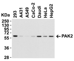
- Western Blot Validation in Human Cell Lines. Loading: 15 µg of lysates per lane. Antibodies: PAK2 Polyclonal Antibody (Product # PA5-20254) (1 µg/mL), 1h incubation at RT in 0.05 NFDM/TBST. Secondary: Goat anti-rabbit IgG HRP conjugate at 1:10,000 dilution.

- Submitted by
- Invitrogen Antibodies (provider)
- Main image

- Experimental details
- Western Blot Validation in Jurkat Lysate. Loading: 15 µg of lysates per lane. Antibodies: PAK2 Polyclonal Antibody (Product # PA5-20254) (A: 0.5 µg/mL, B: 1 µg/mL and C: 2 µg/mL), 1h incubation at RT in 0.05 NFDM/TBST. Secondary: Goat anti-rabbit IgG HRP conjugate at 1:10,000 dilution.

- Submitted by
- Invitrogen Antibodies (provider)
- Main image

- Experimental details
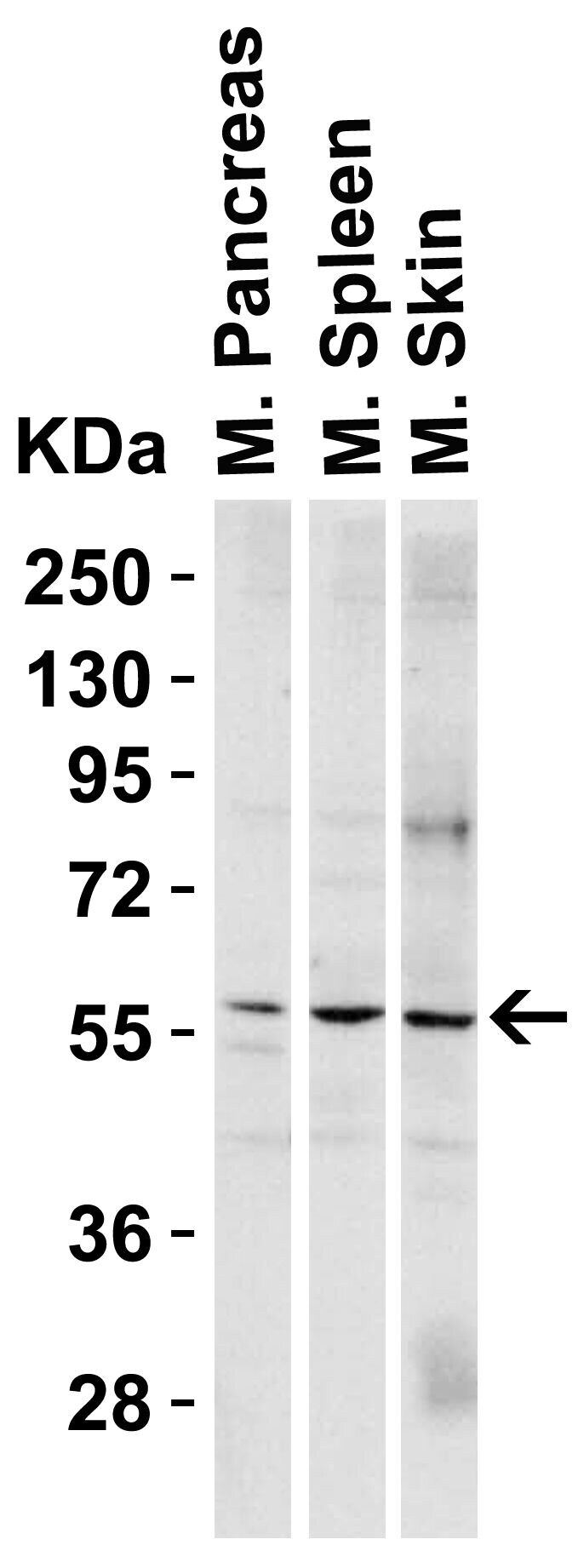
- Western Blot Validation in Mouse Tissues. Loading: 15 µg of lysates per lane. Antibodies: PAK2 Polyclonal Antibody (Product # PA5-20254) (1 µg/mL per lane), 1h incubation at RT in 0.05 NFDM/TBST. Secondary: Goat anti-rabbit IgG HRP conjugate at 1:10,000 dilution.
- Submitted by
- Invitrogen Antibodies (provider)
- Main image
- Experimental details
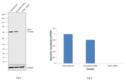
- Knockdown of PAK2 was achieved by transfecting HeLa cells with PAK2 specific siRNAs (Silencer® select Product # S10023). Western blot analysis (Fig. a) was performed using modified membrane enriched cell extracts from the PAK2 knockdown cells (lane 3), non-specific scrambled siRNA transfected cells (lane 2) and untransfected cells (lane 1). The blot was probed with PAK2 Polyclonal Antibody (Product # PA5-20254, 1 µg/mL) and Goat anti-Rabbit IgG (H+L) Superclonal™ Secondary Antibody, HRP conjugate (Product # A27036, 0.25 µg/mL, 1:4000 dilution). Densitometric analysis of this western blot is shown in histogram (Fig b). Decrease in signal upon siRNA mediated knock down confirms that antibody is specific to Cyclin D1.

Supportive validation
- Submitted by
- Invitrogen Antibodies (provider)
- Main image
- Experimental details
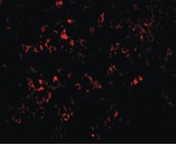
- Immunofluorescent analysis of mouse spleen tissue using a PAK2 polyclonal antibody (Product # PA5-20254) at a 20 µg/mL dilution.

Supportive validation
- Submitted by
- Invitrogen Antibodies (provider)
- Main image

- Experimental details
- Immunofluorescent analysis of 4% paraformaldehyde-fixed mouse spleen tissue labeling PAK2 with PAK2 Polyclonal Antibody (Product # PA5-20254) at 20 µg/mL, followed by goat anti-rabbit IgG secondary antibody at 1:500 dilution (red).

- Submitted by
- Invitrogen Antibodies (provider)
- Main image

- Experimental details
- Immunohistochemical analysis of paraffin-embedded mouse kidney tissue using PAK2 Polyclonal Antibody (Product # PA5-20254) at 10 µg/mL. Tissue was fixed with formaldehyde and blocked with 0.1 serum for 1 h at RT; antigen retrieval was by heat mediation with a citrate buffer (pH6). Samples were incubated with primary antibody overnight at 4˚C. A goat anti-rabbit IgG H&L (HRP) at 1/250 was used as secondary. Counter stained with Hematoxylin.
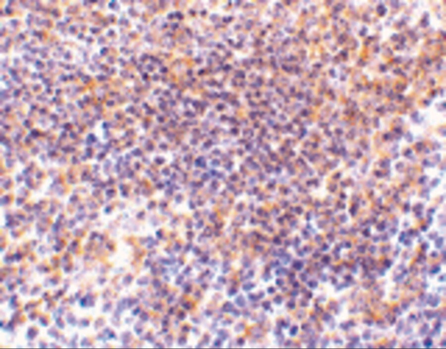
Supportive validation
- Submitted by
- Invitrogen Antibodies (provider)
- Main image

- Experimental details
- NULL
