 Explore
 Validate
 Learn
Learn Western blot
 Immunocytochemistry
Antibody data
- Antibody Data
- Antigen structure
- References [3]
- Comments [0]
- Validations
- Western blot [5]
- Immunocytochemistry [2]
- Immunoprecipitation [1]
- Immunohistochemistry [4]
Submit
Validation data
Reference
Comment
Report error
- Product number
- GTX124213 - Provider product page

- Provider
- GeneTex
- Proper citation
- GeneTex Cat#GTX124213, RRID:AB_11171102
- Product name
- beta Actin antibody
- Antibody type
- Polyclonal
- Reactivity
- Human, Mouse, Rat
- Host
- Rabbit
Submitted references Lymphotoxin β receptor activation promotes mRNA expression of RelA and pro-inflammatory cytokines TNFα and IL-1β in bladder cancer cells.
Proteomics study reveals that the dysregulation of focal adhesion and ribosome contribute to early pregnancy loss.
Sophoricoside fails the embryo implantation by compromising the uterine endometrial receptivity at implantation "window" of pregnant mice.
Shen M, Zhou L, Zhou P, Zhou W, Lin X
Molecular medicine reports 2017 Jul;16(1):937-942
Molecular medicine reports 2017 Jul;16(1):937-942
Proteomics study reveals that the dysregulation of focal adhesion and ribosome contribute to early pregnancy loss.
Xin L, Xu B, Ma L, Hou Q, Ye M, Meng S, Ding X, Ge W
Proteomics. Clinical applications 2016 May;10(5):554-63
Proteomics. Clinical applications 2016 May;10(5):554-63
Sophoricoside fails the embryo implantation by compromising the uterine endometrial receptivity at implantation "window" of pregnant mice.
Zhou J, Qu C, Sun Q, Wu L, Liu Y, Yang Z, Zhang J
Chemico-biological interactions 2014 Aug 5;219:57-63
Chemico-biological interactions 2014 Aug 5;219:57-63
No comments: Submit comment
Enhanced validation
Supportive validation
- Submitted by
- GeneTex (provider)
- Enhanced method
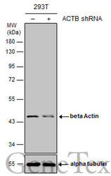
- Genetic validation
- Main image
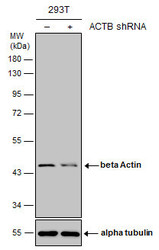
- Experimental details
- Non-transfected (¡V) and transfected (+) 293T whole cell extracts (10 ?g) were separated by 10% SDS-PAGE, and the membrane was blotted with beta Actin antibody (GTX124213) diluted at 1:15000.
Supportive validation
- Submitted by
- GeneTex (provider)
- Main image
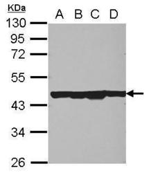
- Experimental details
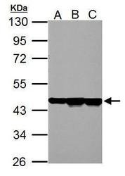
- Sample (30 ug of whole cell lysate) A: 293T B: A431 C: HeLa D: HepG2 10% SDS PAGE GTX124213 diluted at 1:10000
- Submitted by
- GeneTex (provider)
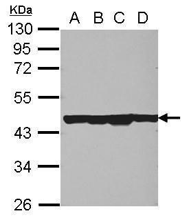
- Main image
- Experimental details
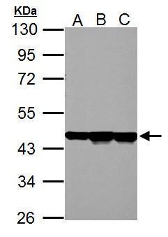
- Sample (30 ug of whole cell lysate) A: NIH-3T3 B: JC C: BCL-1 10% SDS PAGE GTX124213 diluted at 1:10000
- Submitted by
- GeneTex (provider)
- Main image
- Experimental details
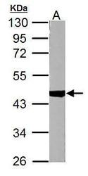
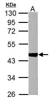
- Sample (30 ug of whole cell lysate) A: PC-12 10% SDS PAGE GTX124213 diluted at 1:10000
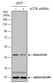
- Submitted by
- GeneTex (provider)
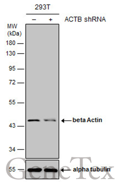
- Main image
- Experimental details
- Non-transfected (¡V) and transfected (+) 293T whole cell extracts (10 ?g) were separated by 10% SDS-PAGE, and the membrane was blotted with beta Actin antibody (GTX124213) diluted at 1:15000.
Supportive validation
- Submitted by
- GeneTex (provider)
- Main image

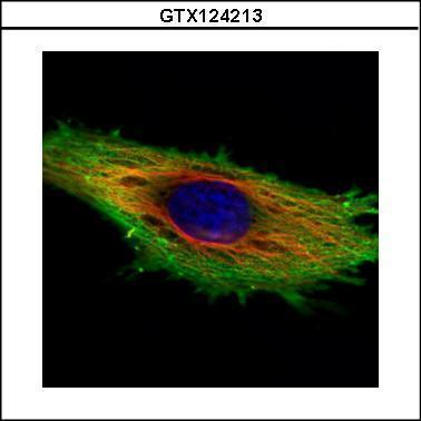
- Experimental details
- Confocal immunofluorescence analysis (Olympus FV10i) of methanol-fixed HeLa, using beta Actin(GTX124213) antibody (Green) at 1:500 dilution. Alpha-tubulin filaments were labeled with GTX11304 (Red) at 1:2000.
- Submitted by
- GeneTex (provider)
- Main image
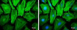
- Experimental details
- beta Actin antibody detects beta Actin protein at cytoskeleton by immunofluorescent analysis.Sample: HeLa cells were fixed in ice-cold MeOH for 5 min.Green: beta Actin protein stained by beta Actin antibody (GTX124213) diluted at 1:500.Blue: Hoechst 33342 staining.Scale bar = 10 £gm.

Supportive validation
- Submitted by
- GeneTex (provider)
- Main image
- Experimental details
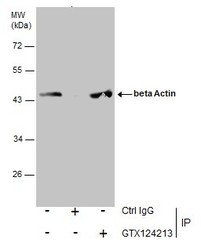
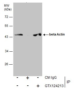
- Immunoprecipitation of beta Actin protein from A431 whole cell extracts using 5 £gg of beta Actin antibody (GTX124213).Western blot analysis was performed using beta Actin antibody (GTX124213).EasyBlot anti-Rabbit IgG (GTX221666-01) was used as a secondary reagent.
Supportive validation
- Submitted by
- GeneTex (provider)
- Main image

- Experimental details
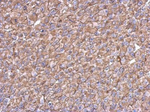
- Immunohistochemical analysis of paraffin-embedded H1299 xenograft, using beta Actin(GTX124213) antibody at 1:500 dilution.
- Submitted by
- GeneTex (provider)
- Main image

- Experimental details
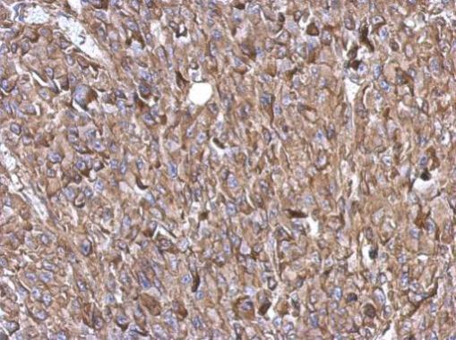
- Immunohistochemical analysis of paraffin-embedded RT2 xenograft, using beta Actin(GTX124213) antibody at 1:500 dilution.
- Submitted by
- GeneTex (provider)
- Main image
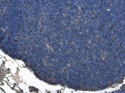
- Experimental details
- Beta Actin antibody detects beta Actin protein at cytoplasm in rat thymus gland by immunohistochemical analysis. Sample: Paraffin-embedded rat thymus gland. Beta Actin antibody (GTX124213) diluted at 1:500.

- Submitted by
- GeneTex (provider)
- Main image
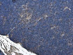
- Experimental details
- Beta Actin antibody detects beta Actin protein at cytoplasm in rat thymus gland by immunohistochemical analysis. Sample: Paraffin-embedded rat thymus gland. Beta Actin antibody (GTX124213) diluted at 1:500.
