Explore
 Validate
 Learn
Learn Western blot
Antibody data
- Antibody Data
- Antigen structure
- References [5]
- Comments [0]
- Validations
- Western blot [1]
- Immunohistochemistry [3]
- Other assay [1]
Submit
Validation data
Reference
Comment
Report error
- Product number
- MA5-17612 - Provider product page

- Provider
- Invitrogen Antibodies
- Product name
- Syntaxin 1 Monoclonal Antibody (SP6)
- Antibody type
- Monoclonal
- Antigen
- Other
- Description
- MA5-17612 targets Syntaxin-1 in ELISA, FACS, and WB applications and shows reactivity with Human, Mouse, and Rat samples.
- Antibody clone number
- SP6
- Concentration
- 1 mg/mL
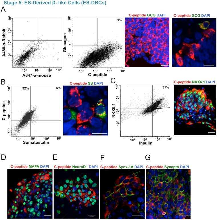
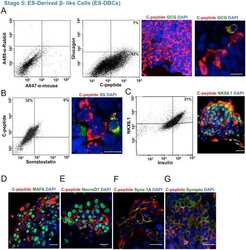
Submitted references An Abbreviated Protocol for In Vitro Generation of Functional Human Embryonic Stem Cell-Derived Beta-Like Cells.
Histone deacetylase expression in white matter oligodendrocytes after stroke.
DNA damage signaling regulates age-dependent proliferative capacity of quiescent inner ear supporting cells.
Intrahepatic myeloid-cell aggregates enable local proliferation of CD8(+) T cells and successful immunotherapy against chronic viral liver infection.
Compton scattering by internal shields based on melanin-containing mushrooms provides protection of gastrointestinal tract from ionizing radiation.
Massumi M, Pourasgari F, Nalla A, Batchuluun B, Nagy K, Neely E, Gull R, Nagy A, Wheeler MB
PloS one 2016;11(10):e0164457
PloS one 2016;11(10):e0164457
Histone deacetylase expression in white matter oligodendrocytes after stroke.
Kassis H, Chopp M, Liu XS, Shehadah A, Roberts C, Zhang ZG
Neurochemistry international 2014 Nov;77:17-23
Neurochemistry international 2014 Nov;77:17-23
DNA damage signaling regulates age-dependent proliferative capacity of quiescent inner ear supporting cells.
Laos M, Anttonen T, Kirjavainen A, af Hällström T, Laiho M, Pirvola U
Aging 2014 Jun;6(6):496-510
Aging 2014 Jun;6(6):496-510
Intrahepatic myeloid-cell aggregates enable local proliferation of CD8(+) T cells and successful immunotherapy against chronic viral liver infection.
Huang LR, Wohlleber D, Reisinger F, Jenne CN, Cheng RL, Abdullah Z, Schildberg FA, Odenthal M, Dienes HP, van Rooijen N, Schmitt E, Garbi N, Croft M, Kurts C, Kubes P, Protzer U, Heikenwalder M, Knolle PA
Nature immunology 2013 Jun;14(6):574-83
Nature immunology 2013 Jun;14(6):574-83
Compton scattering by internal shields based on melanin-containing mushrooms provides protection of gastrointestinal tract from ionizing radiation.
Revskaya E, Chu P, Howell RC, Schweitzer AD, Bryan RA, Harris M, Gerfen G, Jiang Z, Jandl T, Kim K, Ting LM, Sellers RS, Dadachova E, Casadevall A
Cancer biotherapy & radiopharmaceuticals 2012 Nov;27(9):570-6
Cancer biotherapy & radiopharmaceuticals 2012 Nov;27(9):570-6
No comments: Submit comment
Supportive validation
- Submitted by
- Invitrogen Antibodies (provider)
- Main image
- Experimental details
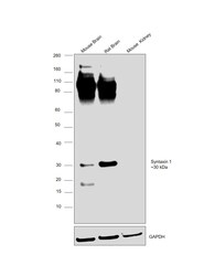
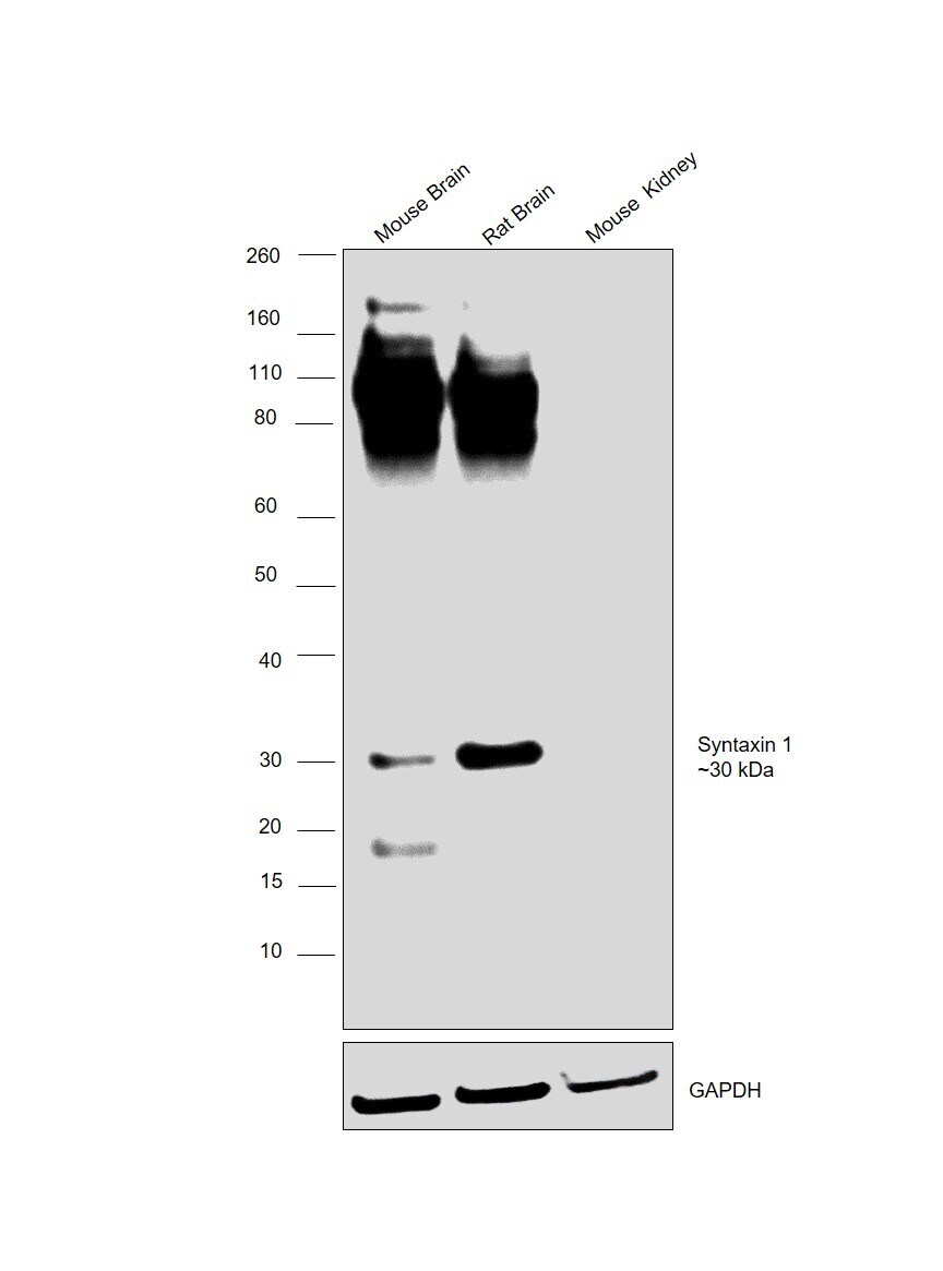
- Western blot was performed using Anti-Syntaxin 1 Monoclonal Antibody (SP6)(Product # MA5-17612) and a 30 kDa band corresponding to Syntaxin 1 was observed across tissues tested. Tissue extracts (30 µg lysate) of Mouse Brain (Lane 1), Rat Brain (Lane 2) and Mouse Kidney (Lane 3) were electrophoresed using NuPAGE™ 4-12% Bis-Tris Protein Gel (Product # NP0321BOX). Resolved proteins were then transferred onto a Nitrocellulose membrane (Product # LC2001) by iBlot® 2 Dry Blotting System (Product # IB21001). The blot was probed with the primary antibody (1:1000 dilution) and detected by chemiluminescence with Goat anti-Mouse IgG (H+L) Superclonal™ Recombinant Secondary Antibody, HRP (Product # A28177,1:4000 dilution) using the iBright FL 1000 (Product # A32752). Chemiluminescent detection was performed using Novex® ECL Chemiluminescent Substrate Reagent Kit (Product # WP20005).Brain is a high expressing model for this protein and Kidney is a low expressing model.
Supportive validation
- Submitted by
- Invitrogen Antibodies (provider)
- Main image
- Experimental details
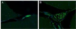
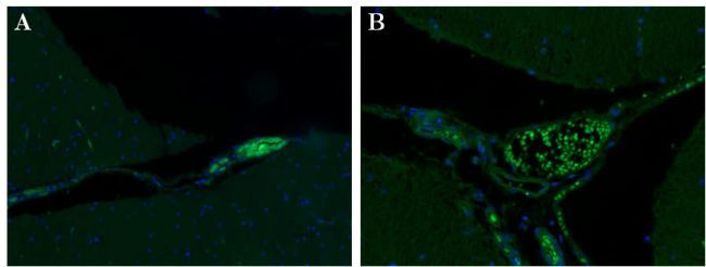
- Immunohistochemical staining of paraformaldehyde-fixed paraffin-embedded human cerebellum sections at 200x (A) and 400x (B) magnification with DAPI counterstain. Primary antibody used was a Syntaxin-1 monoclonal antibody (clone SP6) at a 1:500 dilution, followed by secondary antibody was a 488 fluorescent conjugated Goat anti-mouse Ig at a 1:500 dilution and Normal Goat Serum (blocking)
- Submitted by
- Invitrogen Antibodies (provider)
- Main image
- Experimental details
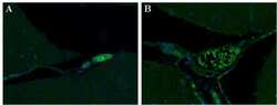
- Immunohistochemical staining of paraformaldehyde-fixed paraffin-embedded human cerebellum sections at 200x (A) and 400x (B) magnification with DAPI counterstain. Primary antibody used was a Syntaxin-1 monoclonal antibody (clone SP6) at a 1:500 dilution, followed by secondary antibody was a 488 fluorescent conjugated Goat anti-mouse Ig at a 1:500 dilution and Normal Goat Serum (blocking)

- Submitted by
- Invitrogen Antibodies (provider)
- Main image
- Experimental details
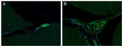
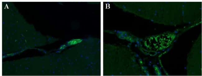
- Immunohistochemical staining of paraformaldehyde-fixed paraffin-embedded human cerebellum sections at 200x (A) and 400x (B) magnification with DAPI counterstain. Primary antibody used was a Syntaxin-1 monoclonal antibody (clone SP6) at a 1:500 dilution, followed by secondary antibody was a 488 fluorescent conjugated Goat anti-mouse Ig at a 1:500 dilution and Normal Goat Serum (blocking)
Supportive validation
- Submitted by
- Invitrogen Antibodies (provider)
- Main image
- Experimental details
- NULL