Explore
 Validate
 Learn
Learn Western blot
 Immunoprecipitation
Antibody data
- Antibody Data
- Antigen structure
- References [37]
- Comments [0]
- Validations
- Western blot [4]
- Immunocytochemistry [1]
- Immunohistochemistry [1]
- Other assay [11]
Submit
Validation data
Reference
Comment
Report error
- Product number
- 33-7100 - Provider product page

- Provider
- Invitrogen Antibodies
- Product name
- MDM2 Monoclonal Antibody (IF2)
- Antibody type
- Monoclonal
- Antigen
- Synthetic peptide
- Description
- This antibody recognizes the ~90 kDa (apparent MW) MDM2 protein. Also recognizes isoforms at ~57 and ~74/76 kDa. The epitope recognized by this antibody is located within amino acids 26-169 of the human protein. Positive controls: OsA-CL, MCF-7, HeLa cell lysates. HOS cells can be used for negative control. Staining of formalin fixed, paraffin embedded tissue requires heat induced epitope retrieval pretreatment.
- Reactivity
- Human
- Host
- Mouse
- Isotype
- IgG
- Antibody clone number
- IF2
- Vial size
- 50 µg
- Concentration
- 0.5 mg/mL
- Storage
- -20° C, Avoid Freeze/Thaw Cycles
Submitted references Decreased DNA Damage and Improved p53 Specificity of RITA Analogs.
The Role of TP53 in Cisplatin Resistance in Mediastinal and Testicular Germ Cell Tumors.
The RNA m6A reader YTHDF2 controls NK cell antitumor and antiviral immunity.
Novel pathogenic alterations in pediatric and adult desmoid-type fibromatosis - A systematic analysis of 204 cases.
E2F-Family Members Engage the PIDDosome to Limit Hepatocyte Ploidy in Liver Development and Regeneration.
Co-expression of MDM2 and CDK4 in transformed human mesenchymal stem cells causes high-grade sarcoma with a dedifferentiated liposarcoma-like morphology.
FoxO3a as a Positive Prognostic Marker and a Therapeutic Target in Tamoxifen-Resistant Breast Cancer.
Establishment and Characterization of Histologically and Molecularly Stable Soft-tissue Sarcoma Xenograft Models for Biological Studies and Preclinical Drug Testing.
A rare case of atypical/anaplastic meningioma with MDM2 amplification.
A New Patient-Derived Metastatic Glioblastoma Cell Line: Characterisation and Response to Sodium Selenite Anticancer Agent.
Association of MDM2 expression with shorter progression-free survival and overall survival in patients with advanced pancreatic cancer treated with gemcitabine-based chemotherapy.
Oridonin synergizes with Nutlin-3 in osteosarcoma cells by modulating the levels of multiple Bcl-2 family proteins.
RITA displays anti-tumor activity in medulloblastomas independent of TP53 status.
MDM2 and CDK4 amplifications are rare events in salivary duct carcinomas.
Ubiquitin-specific protease 2 decreases p53-dependent apoptosis in cutaneous T-cell lymphoma.
Phosphorylated STAT5 regulates p53 expression via BRCA1/BARD1-NPM1 and MDM2.
Prognostic value of HMGA2, CDK4, and JUN amplification in well-differentiated and dedifferentiated liposarcomas.
Myxoid liposarcoma with heterologous components: dedifferentiation or metaplasia? A FISH-documented and CGH-documented case report.
Targeting CDH17 suppresses tumor progression in gastric cancer by downregulating Wnt/β-catenin signaling.
MDM2 and CDK4 immunohistochemical coexpression in high-grade osteosarcoma: correlation with a dedifferentiated subtype.
Pharmacological activation of the p53 pathway by nutlin-3 exerts anti-tumoral effects in medulloblastomas.
MDM2 and CDK4 immunohistochemistry is a valuable tool in the differential diagnosis of low-grade osteosarcomas and other primary fibro-osseous lesions of the bone.
Well-differentiated liposarcoma with low-grade osteosarcomatous component: an underrecognized variant.
Immunohistochemical analysis of MDM2 and CDK4 distinguishes low-grade osteosarcoma from benign mimics.
Similarity in genetic alterations between paired well-differentiated and dedifferentiated components of dedifferentiated liposarcoma.
Markers aiding the diagnosis of chondroid tumors: an immunohistochemical study including osteonectin, bcl-2, cox-2, actin, calponin, D2-40 (podoplanin), mdm-2, CD117 (c-kit), and YKL-40.
Detection of MDM2 gene amplification or protein expression distinguishes sclerosing mesenteritis and retroperitoneal fibrosis from inflammatory well-differentiated liposarcoma.
Lipoleiomyosarcoma of the rectosigmoid colon: a unique site for a rare variant of liposarcoma.
Immunostaining for peroxisome proliferator gamma distinguishes dedifferentiated liposarcoma from other retroperitoneal sarcomas.
Sporadic desmoid tumor. An exceptional cause of cystic pancreatic lesion.
Reproducibility of MDM2 and CDK4 staining in soft tissue tumors.
alpha-fetoprotein expression in a dedifferentiated liposarcoma.
MDM2 as a predictor of prostate carcinoma outcome: an analysis of Radiation Therapy Oncology Group Protocol 8610.
Palmar atypical lipomatous tumour with spindle cell features (well-differentiated spindle cell liposarcoma): a rare neoplasm arising in an unusual anatomical location.
Wild-type p53 overexpression and its correlation with MDM2 and p14ARF alterations: an alternative pathway to non-small-cell lung cancer.
Inflammatory malignant fibrous histiocytomas and dedifferentiated liposarcomas: histological review, genomic profile, and MDM2 and CDK4 status favour a single entity.
Most malignant fibrous histiocytomas developed in the retroperitoneum are dedifferentiated liposarcomas: a review of 25 cases initially diagnosed as malignant fibrous histiocytoma.
Zhan Y, Zhou X, Peuget S, Singh M, Peyser BD, Fan Z, Selivanova G
Molecular cancer therapeutics 2022 Oct 7;21(10):1524-1534
Molecular cancer therapeutics 2022 Oct 7;21(10):1524-1534
The Role of TP53 in Cisplatin Resistance in Mediastinal and Testicular Germ Cell Tumors.
Timmerman DM, Eleveld TF, Gillis AJM, Friedrichs CC, Hillenius S, Remmers TL, Sriram S, Looijenga LHJ
International journal of molecular sciences 2021 Oct 29;22(21)
International journal of molecular sciences 2021 Oct 29;22(21)
The RNA m6A reader YTHDF2 controls NK cell antitumor and antiviral immunity.
Ma S, Yan J, Barr T, Zhang J, Chen Z, Wang LS, Sun JC, Chen J, Caligiuri MA, Yu J
The Journal of experimental medicine 2021 Aug 2;218(8)
The Journal of experimental medicine 2021 Aug 2;218(8)
Novel pathogenic alterations in pediatric and adult desmoid-type fibromatosis - A systematic analysis of 204 cases.
Trautmann M, Rehkämper J, Gevensleben H, Becker J, Wardelmann E, Hartmann W, Grünewald I, Huss S
Scientific reports 2020 Feb 25;10(1):3368
Scientific reports 2020 Feb 25;10(1):3368
E2F-Family Members Engage the PIDDosome to Limit Hepatocyte Ploidy in Liver Development and Regeneration.
Sladky VC, Knapp K, Soratroi C, Heppke J, Eichin F, Rocamora-Reverte L, Szabo TG, Bongiovanni L, Westendorp B, Moreno E, van Liere EA, Bakker B, Spierings DCJ, Wardenaar R, Pereyra D, Starlinger P, Schultze S, Trauner M, Stojakovic T, Scharnagl H, Fava LL, Foijer F, de Bruin A, Villunger A
Developmental cell 2020 Feb 10;52(3):335-349.e7
Developmental cell 2020 Feb 10;52(3):335-349.e7
Co-expression of MDM2 and CDK4 in transformed human mesenchymal stem cells causes high-grade sarcoma with a dedifferentiated liposarcoma-like morphology.
Kim YJ, Kim M, Park HK, Yu DB, Jung K, Song K, Choi YL
Laboratory investigation; a journal of technical methods and pathology 2019 Sep;99(9):1309-1320
Laboratory investigation; a journal of technical methods and pathology 2019 Sep;99(9):1309-1320
FoxO3a as a Positive Prognostic Marker and a Therapeutic Target in Tamoxifen-Resistant Breast Cancer.
Pellegrino M, Rizza P, Donà A, Nigro A, Ricci E, Fiorillo M, Perrotta I, Lanzino M, Giordano C, Bonofiglio D, Bruno R, Sotgia F, Lisanti MP, Sisci D, Morelli C
Cancers 2019 Nov 25;11(12)
Cancers 2019 Nov 25;11(12)
Establishment and Characterization of Histologically and Molecularly Stable Soft-tissue Sarcoma Xenograft Models for Biological Studies and Preclinical Drug Testing.
Cornillie J, Wozniak A, Li H, Wang Y, Boeckx B, Gebreyohannes YK, Wellens J, Vanleeuw U, Hompes D, Stas M, Sinnaeve F, Wafa H, Lambrechts D, Debiec-Rychter M, Sciot R, Schöffski P
Molecular cancer therapeutics 2019 Jun;18(6):1168-1178
Molecular cancer therapeutics 2019 Jun;18(6):1168-1178
A rare case of atypical/anaplastic meningioma with MDM2 amplification.
Wylleman R, Debiec-Rychter M, Sciot R
Rare tumors 2018;10:2036361318779511
Rare tumors 2018;10:2036361318779511
A New Patient-Derived Metastatic Glioblastoma Cell Line: Characterisation and Response to Sodium Selenite Anticancer Agent.
Berthier S, Larrouquère L, Champelovier P, Col E, Lefebvre C, Cottet-Rouselle C, Arnaud J, Garrel C, Laporte F, Boutonnat J, Faure P, Hazane-Puch F
Cancers 2018 Dec 21;11(1)
Cancers 2018 Dec 21;11(1)
Association of MDM2 expression with shorter progression-free survival and overall survival in patients with advanced pancreatic cancer treated with gemcitabine-based chemotherapy.
Yang SH, Lee JC, Guo JC, Kuo SH, Tien YW, Kuo TC, Cheng AL, Yeh KH
PloS one 2017;12(7):e0180628
PloS one 2017;12(7):e0180628
Oridonin synergizes with Nutlin-3 in osteosarcoma cells by modulating the levels of multiple Bcl-2 family proteins.
Wang XH, Zhang SF, Bao JT, Liu FY
Tumour biology : the journal of the International Society for Oncodevelopmental Biology and Medicine 2017 Jun;39(6):1010428317701638
Tumour biology : the journal of the International Society for Oncodevelopmental Biology and Medicine 2017 Jun;39(6):1010428317701638
RITA displays anti-tumor activity in medulloblastomas independent of TP53 status.
Gottlieb A, Althoff K, Grunewald L, Thor T, Odersky A, Schulte M, Deubzer HE, Heukamp L, Eggert A, Schramm A, Schulte JH, Künkele A
Oncotarget 2017 Apr 25;8(17):27882-27891
Oncotarget 2017 Apr 25;8(17):27882-27891
MDM2 and CDK4 amplifications are rare events in salivary duct carcinomas.
Grünewald I, Trautmann M, Busch A, Bauer L, Huss S, Schweinshaupt P, Vollbrecht C, Odenthal M, Quaas A, Büttner R, Meyer MF, Beutner D, Hüttenbrink KB, Wardelmann E, Stenner M, Hartmann W
Oncotarget 2016 Nov 15;7(46):75261-75272
Oncotarget 2016 Nov 15;7(46):75261-75272
Ubiquitin-specific protease 2 decreases p53-dependent apoptosis in cutaneous T-cell lymphoma.
Wei T, Biskup E, Gjerdrum LM, Niazi O, Ødum N, Gniadecki R
Oncotarget 2016 Jul 26;7(30):48391-48400
Oncotarget 2016 Jul 26;7(30):48391-48400
Phosphorylated STAT5 regulates p53 expression via BRCA1/BARD1-NPM1 and MDM2.
Ren Z, Aerts JL, Vandenplas H, Wang JA, Gorbenko O, Chen JP, Giron P, Heirman C, Goyvaerts C, Zacksenhaus E, Minden MD, Stambolic V, Breckpot K, De Grève J
Cell death & disease 2016 Dec 22;7(12):e2560
Cell death & disease 2016 Dec 22;7(12):e2560
Prognostic value of HMGA2, CDK4, and JUN amplification in well-differentiated and dedifferentiated liposarcomas.
Saâda-Bouzid E, Burel-Vandenbos F, Ranchère-Vince D, Birtwisle-Peyrottes I, Chetaille B, Bouvier C, Château MC, Peoc'h M, Battistella M, Bazin A, Gal J, Michiels JF, Coindre JM, Pedeutour F, Bianchini L
Modern pathology : an official journal of the United States and Canadian Academy of Pathology, Inc 2015 Nov;28(11):1404-14
Modern pathology : an official journal of the United States and Canadian Academy of Pathology, Inc 2015 Nov;28(11):1404-14
Myxoid liposarcoma with heterologous components: dedifferentiation or metaplasia? A FISH-documented and CGH-documented case report.
Weingertner N, Neuville A, Chibon F, Ray-Coquard I, Marcellin L, Ghnassia JP
Applied immunohistochemistry & molecular morphology : AIMM 2015 Mar;23(3):230-5
Applied immunohistochemistry & molecular morphology : AIMM 2015 Mar;23(3):230-5
Targeting CDH17 suppresses tumor progression in gastric cancer by downregulating Wnt/β-catenin signaling.
Qiu HB, Zhang LY, Ren C, Zeng ZL, Wu WJ, Luo HY, Zhou ZW, Xu RH
PloS one 2013;8(3):e56959
PloS one 2013;8(3):e56959
MDM2 and CDK4 immunohistochemical coexpression in high-grade osteosarcoma: correlation with a dedifferentiated subtype.
Yoshida A, Ushiku T, Motoi T, Beppu Y, Fukayama M, Tsuda H, Shibata T
The American journal of surgical pathology 2012 Mar;36(3):423-31
The American journal of surgical pathology 2012 Mar;36(3):423-31
Pharmacological activation of the p53 pathway by nutlin-3 exerts anti-tumoral effects in medulloblastomas.
Künkele A, De Preter K, Heukamp L, Thor T, Pajtler KW, Hartmann W, Mittelbronn M, Grotzer MA, Deubzer HE, Speleman F, Schramm A, Eggert A, Schulte JH
Neuro-oncology 2012 Jul;14(7):859-69
Neuro-oncology 2012 Jul;14(7):859-69
MDM2 and CDK4 immunohistochemistry is a valuable tool in the differential diagnosis of low-grade osteosarcomas and other primary fibro-osseous lesions of the bone.
Dujardin F, Binh MB, Bouvier C, Gomez-Brouchet A, Larousserie F, Muret Ad, Louis-Brennetot C, Aurias A, Coindre JM, Guillou L, Pedeutour F, Duval H, Collin C, de Pinieux G
Modern pathology : an official journal of the United States and Canadian Academy of Pathology, Inc 2011 May;24(5):624-37
Modern pathology : an official journal of the United States and Canadian Academy of Pathology, Inc 2011 May;24(5):624-37
Well-differentiated liposarcoma with low-grade osteosarcomatous component: an underrecognized variant.
Yoshida A, Ushiku T, Motoi T, Shibata T, Fukayama M, Tsuda H
The American journal of surgical pathology 2010 Sep;34(9):1361-6
The American journal of surgical pathology 2010 Sep;34(9):1361-6
Immunohistochemical analysis of MDM2 and CDK4 distinguishes low-grade osteosarcoma from benign mimics.
Yoshida A, Ushiku T, Motoi T, Shibata T, Beppu Y, Fukayama M, Tsuda H
Modern pathology : an official journal of the United States and Canadian Academy of Pathology, Inc 2010 Sep;23(9):1279-88
Modern pathology : an official journal of the United States and Canadian Academy of Pathology, Inc 2010 Sep;23(9):1279-88
Similarity in genetic alterations between paired well-differentiated and dedifferentiated components of dedifferentiated liposarcoma.
Horvai AE, DeVries S, Roy R, O'Donnell RJ, Waldman F
Modern pathology : an official journal of the United States and Canadian Academy of Pathology, Inc 2009 Nov;22(11):1477-88
Modern pathology : an official journal of the United States and Canadian Academy of Pathology, Inc 2009 Nov;22(11):1477-88
Markers aiding the diagnosis of chondroid tumors: an immunohistochemical study including osteonectin, bcl-2, cox-2, actin, calponin, D2-40 (podoplanin), mdm-2, CD117 (c-kit), and YKL-40.
Daugaard S, Christensen LH, Høgdall E
APMIS : acta pathologica, microbiologica, et immunologica Scandinavica 2009 Jul;117(7):518-25
APMIS : acta pathologica, microbiologica, et immunologica Scandinavica 2009 Jul;117(7):518-25
Detection of MDM2 gene amplification or protein expression distinguishes sclerosing mesenteritis and retroperitoneal fibrosis from inflammatory well-differentiated liposarcoma.
Weaver J, Goldblum JR, Turner S, Tubbs RR, Wang WL, Lazar AJ, Rubin BP
Modern pathology : an official journal of the United States and Canadian Academy of Pathology, Inc 2009 Jan;22(1):66-70
Modern pathology : an official journal of the United States and Canadian Academy of Pathology, Inc 2009 Jan;22(1):66-70
Lipoleiomyosarcoma of the rectosigmoid colon: a unique site for a rare variant of liposarcoma.
Nahal A, Meterissian S
American journal of clinical oncology 2009 Aug;32(4):353-5
American journal of clinical oncology 2009 Aug;32(4):353-5
Immunostaining for peroxisome proliferator gamma distinguishes dedifferentiated liposarcoma from other retroperitoneal sarcomas.
Horvai AE, Schaefer JT, Nakakura EK, O'Donnell RJ
Modern pathology : an official journal of the United States and Canadian Academy of Pathology, Inc 2008 May;21(5):517-24
Modern pathology : an official journal of the United States and Canadian Academy of Pathology, Inc 2008 May;21(5):517-24
Sporadic desmoid tumor. An exceptional cause of cystic pancreatic lesion.
Amiot A, Dokmak S, Sauvanet A, Vilgrain V, Bringuier PP, Scoazec JY, Sastre X, Ruszniewski P, Bedossa P, Couvelard A
JOP : Journal of the pancreas 2008 May 8;9(3):339-45
JOP : Journal of the pancreas 2008 May 8;9(3):339-45
Reproducibility of MDM2 and CDK4 staining in soft tissue tumors.
Binh MB, Garau XS, Guillou L, Aurias A, Coindre JM
American journal of clinical pathology 2006 May;125(5):693-7
American journal of clinical pathology 2006 May;125(5):693-7
alpha-fetoprotein expression in a dedifferentiated liposarcoma.
Bosco M, Allia E, Coindre JM, Odasso C, Pagani A, Pacchioni D
Virchows Archiv : an international journal of pathology 2006 Apr;448(4):517-20
Virchows Archiv : an international journal of pathology 2006 Apr;448(4):517-20
MDM2 as a predictor of prostate carcinoma outcome: an analysis of Radiation Therapy Oncology Group Protocol 8610.
Khor LY, Desilvio M, Al-Saleem T, Hammond ME, Grignon DJ, Sause W, Pilepich M, Okunieff P, Sandler H, Pollack A, Radiation Therapy Oncology Group
Cancer 2005 Sep 1;104(5):962-7
Cancer 2005 Sep 1;104(5):962-7
Palmar atypical lipomatous tumour with spindle cell features (well-differentiated spindle cell liposarcoma): a rare neoplasm arising in an unusual anatomical location.
Mentzel T, Toennissen J, Rütten A, Schaller J
Virchows Archiv : an international journal of pathology 2005 Mar;446(3):300-4
Virchows Archiv : an international journal of pathology 2005 Mar;446(3):300-4
Wild-type p53 overexpression and its correlation with MDM2 and p14ARF alterations: an alternative pathway to non-small-cell lung cancer.
Wang YC, Lin RK, Tan YH, Chen JT, Chen CY, Wang YC
Journal of clinical oncology : official journal of the American Society of Clinical Oncology 2005 Jan 1;23(1):154-64
Journal of clinical oncology : official journal of the American Society of Clinical Oncology 2005 Jan 1;23(1):154-64
Inflammatory malignant fibrous histiocytomas and dedifferentiated liposarcomas: histological review, genomic profile, and MDM2 and CDK4 status favour a single entity.
Coindre JM, Hostein I, Maire G, Derré J, Guillou L, Leroux A, Ghnassia JP, Collin F, Pedeutour F, Aurias A
The Journal of pathology 2004 Jul;203(3):822-30
The Journal of pathology 2004 Jul;203(3):822-30
Most malignant fibrous histiocytomas developed in the retroperitoneum are dedifferentiated liposarcomas: a review of 25 cases initially diagnosed as malignant fibrous histiocytoma.
Coindre JM, Mariani O, Chibon F, Mairal A, De Saint Aubain Somerhausen N, Favre-Guillevin E, Bui NB, Stoeckle E, Hostein I, Aurias A
Modern pathology : an official journal of the United States and Canadian Academy of Pathology, Inc 2003 Mar;16(3):256-62
Modern pathology : an official journal of the United States and Canadian Academy of Pathology, Inc 2003 Mar;16(3):256-62
No comments: Submit comment
Supportive validation
- Submitted by
- Invitrogen Antibodies (provider)
- Main image

- Experimental details
- Western blot analysis of MDM2 was performed by loading 30 µg of the indicated whole cell lysates and 5 µL of PageRuler Plus Prestained Protein Ladder (Product # 26619) per well onto a Novex 4-20% Tris-Glycine polyacrylamide gel (Product # WT4202BOX ). Proteins were transferred to a nitrocellulose membrane using the G2 Blotter (Product # 62288), and blocked with 5% Milk in TBST for 1 hour at room temperature. MDM2 was detected at ~57, ~75, and ~90 kDa using a MDM2 monoclonal antibody (Product # 33-7100) at a dilution of 2.5 µg/mL in 5% Milk in TBST overnight at 4C on a rocking platform, followed by a Goat anti-Mouse IgG (H+L) Superclonal Secondary Antibody, HRP conjugate (Product # A28177) at a dilution of 1:1000 for at least 30 minutes at room temperature. Chemiluminescent detection was performed using SuperSignal Pico substrate (Product # 34078) and the myECL Imager (Product # 62236).

- Submitted by
- Invitrogen Antibodies (provider)
- Main image

- Experimental details
- Knockdown of MDM2 was achieved by transfecting U-2 OS with MDM2 specific siRNAs (Silencer® select Product # S8628, S8629). Western blot analysis (Fig. a) was performed using Whole cell extracts from the MDM2 knockdown cells (lane 3), non-targeting scrambled siRNA transfected cells (lane 2) and untransfected cells (lane 1). The blot was probed with MDM2 Monoclonal Antibody (IF2) (Product # 33-7100, 1 µg/mL ) and Goat anti-Mouse IgG (H+L) Superclonal™ Recombinant Secondary Antibody, HRP (Product # A28177, 1:4000 dilution). Densitometric analysis of this western blot is shown in histogram (Fig. b). Decrease in signal upon siRNA mediated knockdown confirms that antibody is specific to MDM2.

- Submitted by
- Invitrogen Antibodies (provider)
- Main image

- Experimental details
- Western blot was performed using Anti-MDM2 Monoclonal Antibody (IF2) (Product # 33-7100) and a 75 kDa band along with 55 kDa band, both corresponding to MDM2 was observed across cell lines tested. Whole cell extracts (30 µg lysate) of U-2 OS (Lane 1) and A549 (Lane 2) were electrophoresed using NuPAGE™ 4-12% Bis-Tris Protein Gel (Product # NP0322BOX). Resolved proteins were then transferred onto a Nitrocellulose membrane (Product # IB23001) by iBlot® 2 Dry Blotting System (Product # IB21001). The blot was probed with the primary antibody (1 µg/mL) and detected by chemiluminescence with Goat anti-Mouse IgG (H+L) Superclonal™ Recombinant Secondary Antibody, HRP (Product # A28177, 1:4000 dilution) using the iBright FL 1000 (Product # A32752). Chemiluminescent detection was performed using SuperSignal™ West Dura Extended Duration Substrate (Product # 34076).

Supportive validation
- Submitted by
- Invitrogen Antibodies (provider)
- Main image

- Experimental details
- Immunofluorescence analysis of MDM2 in subconfluent U2OS cells. The cells were fixed with 4% paraformaldehyde for 15 minutes, permeabilized with 0.1% Triton X-100 for 15 minutes, and blocked with 3% BSA for 15 minutes at room temperature. The cells were probed with a MDM2 Mouse Monoclonal Antibody (Product # 33-7100) at 1.5 µg/mL for 1 hour at room temperature and then labeled with a Goat anti-Mouse IgG (H+L) Superclonal Secondary Antibody, Alexa Fluor 488 conjugate (Product # A28175) at a dilution of 1:400 for 30 minutes at room temperature (Panel a: green). Nuclei (Panel b: blue) were stained with Hoechst Dye. F-actin (Panel c: red) was stained with DyLight 554 Phalloidin (Product # 21834). Panel d is a merged image showing predominantly nuclear localization. Panel e shows no primary antibody control. The images were captured at 20X magnification.

Supportive validation
- Submitted by
- Invitrogen Antibodies (provider)
- Main image

- Experimental details
- Immunohistochemistry analysis of MDM2 showing staining in the cytoplasm and nucleus of paraffin-embedded human breast carcinoma (right) compared to a negative control without primary antibody (left). To expose target proteins, antigen retrieval was performed using 10mM sodium citrate (pH 6.0) and heated in a 95C water bath for 20 minutes. Following antigen retrieval, tissues were blocked in 10% goat serum in PBS for 30 minutes at room temperature and quenched with Peroxide Suppressor (Product # 35000) for 30 minutes. Tissues were then probed with a MDM2 monoclonal antibody (Product # 33-7100) at a dilution of 40 µg/mL in blocking buffer for 1 hour at room temperature. Tissues were washed extensively in PBST and detection was performed using the SuperPicture HRP Polymer Detection Kit (Product # 87-8963) and DAB substrate (Product # 34002). Tissues were counterstained with hematoxylin (Product # TA-125-MH) and dehydrated with ethanol and xylene to prep for mounting.

Supportive validation
- Submitted by
- Invitrogen Antibodies (provider)
- Main image

- Experimental details
- Figure 1. (a) Hematoxylin and eosin stain (40x magnification) from the first, atypicalmeningioma showed a highly cellular tumor with large nuclei and prominentnucleoli, and an increased mitotic count. (b) The tumor showed a varyingexpression pattern for EMA (20x magnification). (c) SSTR2A showed diffuseand strong membranous and cytoplasmic positivity (20x magnification). (d)MDM2 amplification resulted in MDM2 overexpression, since additionalimmunohistochemistry showed nuclear positivity for MDM2 (40xmagnification).

- Submitted by
- Invitrogen Antibodies (provider)
- Main image

- Experimental details
- Figure 2. a) Hematoxylin and eosin stain (40x magnification) from the recurrent,anaplastic meningioma showed a frankly malignant tumor with prominentcytological atypia, high mitotic count, and extensive necrosis. (b) Thetumor showed only partial positivity for EMA (20x magnification). (c) SSTR2Ashowed membranous and cytoplasmic positivity in the majority of the tumor(40x magnification). (d) MDM2 amplification resulted in MDM2 overexpression,since additional immunohistochemistry showed nuclear positivity for MDM2(40x magnification).

- Submitted by
- Invitrogen Antibodies (provider)
- Main image

- Experimental details
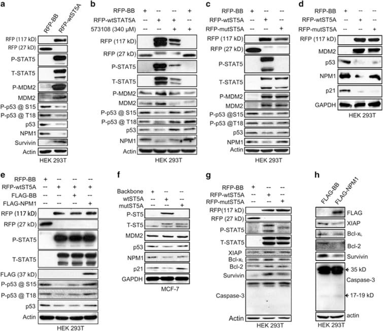
- Figure 5 P-STAT5 regulates p53/MDM2 functions and cell survival through NPM1 protein. ( a ) HEK 293T cells (6 x 10 5 per well) transfected with either RFP-STAT5A or RFP-backbone vectors were harvested at 24 h after transfection, and the cell lysates were subjected to immunoblotting assays for detection of expression levels of p53 and its phosphorylation levels at serine 15 and threonine 18, as well as the expression levels of MDM2 and its phosphorylation level at serine 166. ( b ) Six hours after transfection with either RFP-STAT5A or RFP-backbone vectors, HEK 293T cells (6 x 10 5 per well) were incubated overnight with inhibitor 573108 at a concentration of 340 mu M and then harvested and lysed for immunoblotting assays to examine the expression levels of p53 and MDM2 as well as their relevant phosphorylation levels. ( c and d ) HEK 293T cells (6 x 10 5 per well) were transfected with RFP-STAT5AY694F mutant vector for 24 h, harvested and lysed for immunoblotting assays on the expression levels of MDM2, p53 and p21. ( e ) HEK 293T cells (6 x 10 5 per well) were co-transfected with RFP-wtSTAT5A and FLAG-NPM1 vectors for 24 h, harvested and lysed for immunoblotting assays on the p53 expression levels. ( f ) MCF-7 cells (2 x 10 6 per well) were plated on 10 cm plate on day 0 and then lentivirally transduced with pLVX-IRES-mCherry-wtSTAT5 or pLVX-IRES-mCherry-STAT5Y694F on day 1. The cells were harvested 72 h after the transduction and lysed for immunoblotting assays. ( g ) HEK 2
- Submitted by
- Invitrogen Antibodies (provider)
- Main image

- Experimental details
- Figure 2 USP2 is induced by PUVA and a p53 activator, nutlin3a p53 wt CTCL cell line, MyLa2000, was subjected to PUVA a, b. or 5muM nutlin3a c-d. as shown in Methods. USP2 expression was measured by qPCR (a, c) and western blot (WB) (b, d) 2h-72h after the treatments. For qPCR, data was normalized to GAPDH and expressed as relative units (RU). The experiments were repeated 3 times, unpaired T-test was used to calculate P-value, error-bars, SD, *, P

- Submitted by
- Invitrogen Antibodies (provider)
- Main image

- Experimental details
- Figure 5 USP2 inhibits the cellular actions of p53 through Mdm2 a. USP2 knockdown decreased Mdm2 expression. MyLa2000 cells were transfected with either siRNA control or USP2 siRNA for 24h, followed by treatment with 5muM nutlin3a for 24h. Western blot was used for detecting USP2, Mdm2 and p53 protein level. beta-actin was used as internal control, and relative protein expression levels are reported below the corresponding western blot bands. The experiment was repeated 3 times. Representative data was shown. b. USP2 knockdown increase p53 transcription activity. The expression of p21 was used to indicate p53 transcription activity. qPCR was applied to examine p21 expression in MyLa2000 cells transfected with siUSP2/siRNA with or without nutlin3a treatment. Paired T-test was used to calculate P-value. c. A proposed model for the regulation of USP2. PUVA and nutlin3a activate p53 and promote cell apoptosis. USP2 is induced by PUVA and nutlin3a and increases cell resistance to apoptosis via modulation of Mdm2/p53 interaction, constituting an anti-apoptosis protective loop.

- Submitted by
- Invitrogen Antibodies (provider)
- Main image

- Experimental details
- Figure 2 RITA restores TP53 activity in medulloblastoma cell lines with and without TP53 mutations ( A ) TP53 and CDKN1A protein expression after 48 h of RITA treatment assessed by western blot. Ethanol served as a negative control. wt = wildtype, mut = mutation. ( B, C ) Relative expression of CDKN1A and MDM2 mRNA after 48 h of RITA treatment measured by real-time polymerase chain reaction (PCR). Ethanol served as a negative control. * = P < 0.05 in Student's t-test .

- Submitted by
- Invitrogen Antibodies (provider)
- Main image

- Experimental details
- Figure 5 CDH17 activates multiple signal transduction pathways. A. Expression of beta-catenin, GSK-3beta, p- GSK-3beta, Rb, Cyclin D1were compared between shCDH17 and Mock both in AGS and MKN-45 cells by Western blot analysis. MDM2, p53 and p21 also were evaluated. beta-Actin was used as a loading control. B. TOPflash/FOPflash reporter assay shows that Wnt signaling re-activated after CDH17 restoration in AGS and MKN-45. (* P

- Submitted by
- Invitrogen Antibodies (provider)
- Main image
- Experimental details
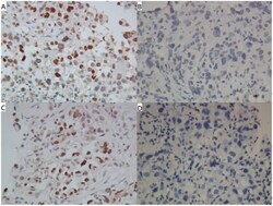
- Fig 1 Representative cases of IHC expression (magnification 400X). Cases of IHC expression with (A) MDM2+, (B) MDM2-, (C) p53+, and (D) p53- were demonstrated. The positive staining was predominantly nuclear for both MDM2 and p53.

- Submitted by
- Invitrogen Antibodies (provider)
- Main image

- Experimental details
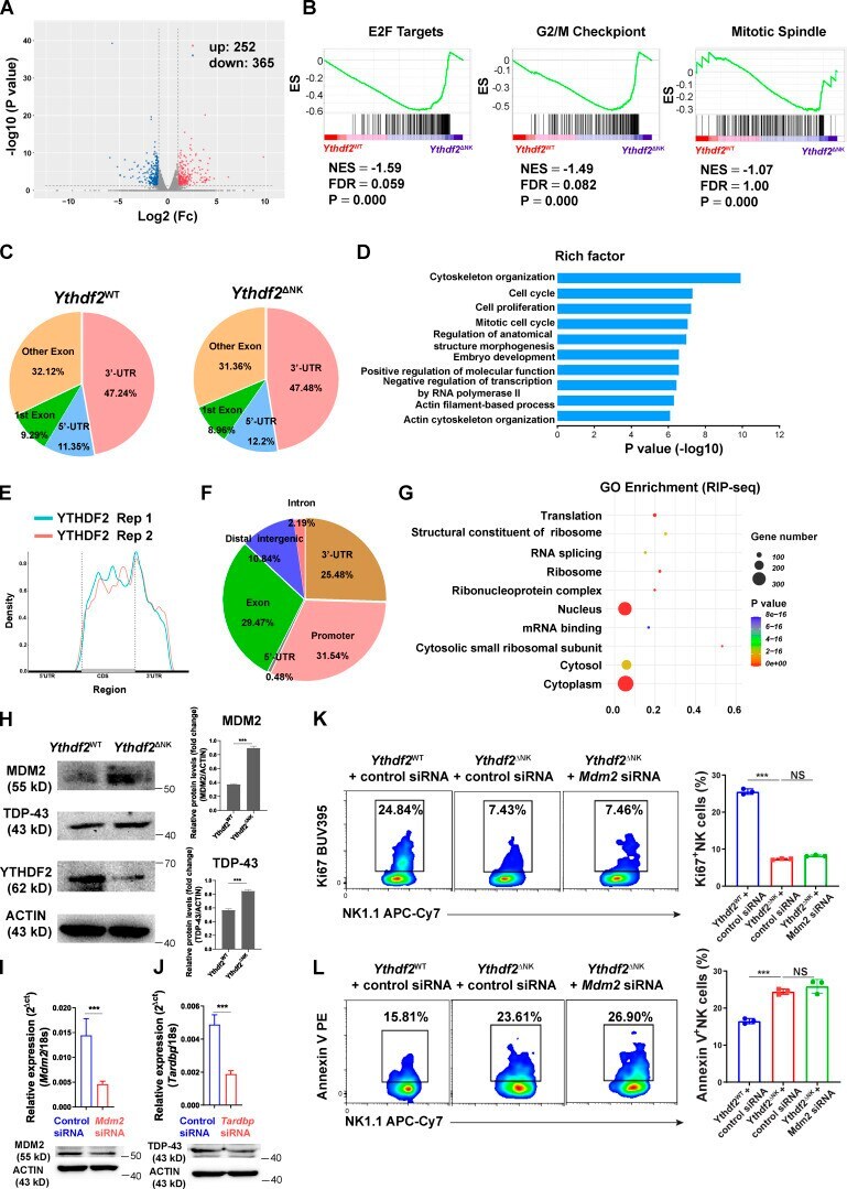
- Figure S5. Transcriptome-wide RNA-seq, m 6 A-seq, and RIP-seq assays in murine NK cells. (A) Volcano plots of differentially expressed genes from RNA-seq. (B) GSEA showing enrichment of E2F targets, G2/M checkpoint, and mitotic spindle hallmark gene sets in Ythdf2 DeltaNK NK cells compared with Ythdf2 WT NK cells. (C) The proportion of m 6 A peak distribution in NK cells from Ythdf2 WT and Ythdf2 DeltaNK mice. (D) GO analysis of transcripts with m 6 A peaks. (E) Density distribution of the YTHDF2-binding sites across mRNA transcriptome from RIP-seq data. (F) The proportion of YTHDF2 binding site distribution from RIP-seq data. (G) Top 10 GO clusters from GO analysis of YTHDF2 target genes from RIP-seq data. (H) Immunoblotting showing the protein levels of MDM2, TDP-43, and YTHDF2 in IL-2-expanded NK cells enriched from the spleen of Ythdf2 WT and Ythdf2 DeltaNK mice. (I and J) qPCR and immunoblotting showing the expression of Mdm2 (I) and Tardbp (J) in NK cells transfected with gene-specific or control siRNA. (K and L) IL-2-expanded NK cells were transfected with Mdm2 -specific siRNA cells under the stimulation of IL-15. 3 d later, cell proliferation and apoptosis were analyzed by Ki67 staining (K) and annexin V staining (L), respectively ( n = 3 per group). Data are shown as mean +- SD and were analyzed by unpaired two-tailed t test (I and J) or one-way ANOVA with Sidak post-test (K and L). Data are representative of at least two independent experiments. ***, P < 0.001. ES,
- Submitted by
- Invitrogen Antibodies (provider)
- Main image

- Experimental details
- Figure 2 Characterization of the cell lines NCCIT and 2102Ep. ( A ) Schematic overview of the hemizygous mutation present in the NCCIT cell line. ( B ) Bar graph displaying the normalized expression (RNA-seq) of MDM2 in the NCCIT and 2102Ep parental and resistant cell lines. ( C ) Western blot showing the protein levels of MDM2 in the NCCIT and 2102Ep parental and resistant cell lines. ( D , E ) Western blot displaying the MDM2 ( D ) and MDM4 ( E ) protein levels after treatment with sublethal cisplatin doses (1 uM) or saline vehicle control. ( F ) Mutational position of TP53 mutations in patients in the MSKCC, J Clin Oncol 2016 data set. The mutation found in NCCIT is highlighted with a blue dot.

- Submitted by
- Invitrogen Antibodies (provider)
- Main image

- Experimental details
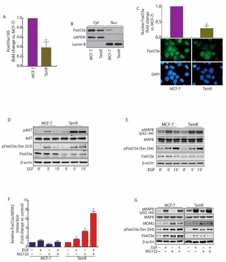
- Figure 1 FoxO3a is downregulated in TamR BBCs. ( A ) FoxO3a transcripts were analyzed by real-time PCR in growing MCF-7 and TamR cells. Each sample was normalized vs. its 18S rRNA content and presented as fold enrichment versus MCF-7. The results represent the mean +- s.d. of three independent experiments. *, p < 0.01 vs. untreated. ( B ) Cytoplasmic and nuclear protein extracts from a duplicate set of cells were subjected to WB (30 mug/lane) to evaluate the subcellular localization of FoxO3a. GAPDH and Lamin B (cytosolic and nuclear markers, respectively) were used as loading controls and to assess the quality of the subcellular protein fractionation. ( C ) Immunostaining of FoxO3a expression and localization (green) in MCF-7 and TamR growing cells; nuclear integrity was visualized by DAPI (blue) (400x magnification) ( D , E ) Comparison between AKT ( D ) and MAPK ( E ) signal transduction pathways in MCF-7 and TamR cells. Cells were starved in PRF-SFM for 16 h and then treated or not with EGF (100 nM). Proteins were analyzed by WB, using the indicated antibodies. ( F ) PLA. MCF-7 and TamR cells were seeded in MW8 (Lab-Tek(tm) Chamber Slide System, Nunc(tm)), left to adhere for 48 h, then starved in PRF-SFM and pre-treated with MG132 (20 muM) or left untreated (-). The next day, EGF (100 nM) was added for 30 min where indicated. Antibodies against FoxO3a and MDM2 were used to detect the active complexes. Captures of the stained FoxO3a/MDM2 complexes were analyzed by ImageJ s