 Explore
 Validate
 Learn
Learn Western blot
 ELISA
ELISAAntibody data
- Antibody Data
- Antigen structure
- References [2]
- Comments [0]
- Validations
- Western blot [1]
- Immunohistochemistry [2]
Submit
Validation data
Reference
Comment
Report error
- Product number
- MAB22441-100 - Provider product page

- Provider
- R&D Systems
- Product name
- Human CEACAM-1/CD66a Antibody
- Antibody type
- Monoclonal
- Description
- Protein A or G purified from hybridoma culture supernatant. Detects human CEACAM-1 in ELISAs and Western blots. In ELISAs and Western blots, no cross-reactivity with recombinant human (rh) CD31, rhICAM-1, -2, -3, recombinant mouse MAdCAM-1, or rhVCAM-1 was observed. In sandwich ELISAs, no cross-reactivity with rhCEACAM-3, rhCEACAM-5, or rhCEACAM-6 was observed.
- Reactivity
- Human
- Host
- Mouse
- Conjugate
- Unconjugated
- Antigen sequence
P13688- Isotype
- IgG
- Antibody clone number
- 283324
- Vial size
- 100 ug
- Storage
- Use a manual defrost freezer and avoid repeated freeze-thaw cycles. 12 months from date of receipt, -20 to -70 °C as supplied. 1 month, 2 to 8 °C under sterile conditions after reconstitution. 6 months, -20 to -70 °C under sterile conditions after reconstitution.
Submitted references Serum CEACAM1 Level Is Associated with Diagnosis and Prognosis in Patients with Osteosarcoma.
AP-1-controlled hepatocyte growth factor activation promotes keratinocyte migration via CEACAM1 and urokinase plasminogen activator/urokinase plasminogen receptor.
Yu H, Yu J, Ren Y, Yang Y, Xiao X
PloS one 2016;11(4):e0153601
PloS one 2016;11(4):e0153601
AP-1-controlled hepatocyte growth factor activation promotes keratinocyte migration via CEACAM1 and urokinase plasminogen activator/urokinase plasminogen receptor.
Schnickmann S, Camacho-Trullio D, Bissinger M, Eils R, Angel P, Schirmacher P, Szabowski A, Breuhahn K
The Journal of investigative dermatology 2009 May;129(5):1140-8
The Journal of investigative dermatology 2009 May;129(5):1140-8
No comments: Submit comment
Supportive validation
- Submitted by
- R&D Systems (provider)
- Main image

- Experimental details
- Detection of Human CEACAM-1/CD66a by Western Blot. Western blot shows lysates of human liver tissue and HepG2 human hepatocellular carcinoma cell line. PVDF membrane was probed with 2 µg/mL of Mouse Anti-Human CEACAM-1/CD66a Monoclonal Antibody (Catalog # MAB22441) followed by HRP-conjugated Anti-Mouse IgG Secondary Antibody (Catalog # HAF018). Specific bands were detected for CEACAM-1/CD66a at approximately 100-150 kDa (as indicated). This experiment was conducted under reducing conditions and using Immunoblot Buffer Group 1.

Supportive validation
- Submitted by
- R&D Systems (provider)
- Main image
- Experimental details
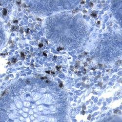
- CEACAM-1/CD66a in Human Colon. CEACAM-1/CD66a was detected in immersion fixed paraffin-embedded sections of human colon using Mouse Anti-Human CEACAM-1/CD66a Monoclonal Antibody (Catalog # MAB22441) at 25 µg/mL overnight at 4 °C. Tissue was stained using the Anti-Mouse HRP-DAB Cell & Tissue Staining Kit (brown; Catalog # CTS002) and counterstained with hematoxylin (blue). Specific labeling was localized to stromal cells. View our protocol for Chromogenic IHC Staining of Paraffin-embedded Tissue Sections.

- Submitted by
- R&D Systems (provider)
- Main image

- Experimental details
- CEACAM-1/CD66a in Human Colon Cancer Tissue. CEACAM-1/CD66a was detected in immersion fixed paraffin-embedded sections of human colon cancer tissue using Mouse Anti-Human CEACAM-1/CD66a Monoclonal Antibody (Catalog # MAB22441) at 25 µg/mL over-night at 4 °C. Tissue was stained using the Anti-Mouse HRP-DAB Cell & Tissue Staining Kit (brown; Catalog # CTS002) and counterstained with hematoxylin (blue). View our protocol for Chromogenic IHC Staining of Paraffin-embedded Tissue Sections.
