Explore
 Validate
 Learn
Learn Western blot
Antibody data
- Antibody Data
- Antigen structure
- References [1]
- Comments [0]
- Validations
- Western blot [4]
- Immunocytochemistry [1]
- Immunohistochemistry [2]
- Other assay [1]
Submit
Validation data
Reference
Comment
Report error
- Product number
- PA5-79677 - Provider product page

- Provider
- Invitrogen Antibodies
- Product name
- MMP10 Polyclonal Antibody
- Antibody type
- Polyclonal
- Antigen
- Recombinant full-length protein
- Description
- Reconstitute with 0.2 mL of distilled water to yield a concentration of 500 µg/mL.
- Reactivity
- Human, Mouse, Rat
- Host
- Rabbit
- Isotype
- IgG
- Vial size
- 100 µg
- Concentration
- 500 µg/mL
- Storage
- -20°C
Submitted references Evidence of a Myenteric Plexus Barrier and Its Macrophage-Dependent Degradation During Murine Colitis: Implications in Enteric Neuroinflammation.
Dora D, Ferenczi S, Stavely R, Toth VE, Varga ZV, Kovacs T, Bodi I, Hotta R, Kovacs KJ, Goldstein AM, Nagy N
Cellular and molecular gastroenterology and hepatology 2021;12(5):1617-1641
Cellular and molecular gastroenterology and hepatology 2021;12(5):1617-1641
No comments: Submit comment
Supportive validation
- Submitted by
- Invitrogen Antibodies (provider)
- Main image

- Experimental details
- Western blot analysis of MMP10 in Lane 1: rat cardiac muscle tissue lysate, Lane 2: mouse cardiac muscle tissue lysate using 50 µg (reducing conditions) per well. Electrophoresis was performed on 5-20% SDS-PAGE gel at 70V (Stacking gel) / 90V (Resolving gel) for 2-3 hours and protein was transferred to a nitrocellulose membrane at 150mA for 50-90 minutes. Sample was blocked with 5% Non-fat Milk/TBS for 1.5 hours at room temperature, incubated with MMP10 polyclonal antibody (Product # PA5-79677) at a dilution of 0.5 µg/mL (overnight at 4°C), followed by goat anti-rabbit IgG-HRP secondary antibody at a dilution of 1:10,000. Signal development was performed using a chemiluminescence (ECL) kit.

- Submitted by
- Invitrogen Antibodies (provider)
- Main image

- Experimental details
- Western blot analysis of MMP10 in, Lane 1: human placenta tissue lysates. Electrophoresis was performed on a 5-20% SDS-PAGE gel at 70V (Stacking gel) / 90V (Resolving gel) for 2-3 hours. The sample well of each lane was loaded with 50 µg of sample under reducing conditions. After Electrophoresis, proteins were transferred to a Nitrocellulose membrane at 150mA for 50-90 minutes. The membrane was blocked with 5% Non-fat Milk/ TBS for 1. 5 hour at RT. The membrane was incubated with MMP10 Polyclonal Antibody (Product # PA5-79677) at 0.5 μg/mL overnight at 4°C, then washed with TBS-0. 1% Tween 3 times with 5 minutes each and probed with a goat anti-rabbit IgG-HRP secondary antibody at a dilution of 1:10000 for 1. 5 hour at RT. The signal is developed using an Enhanced Chemiluminescent detection (ECL) kit. A specific band was detected for MMP10 at approximately 54KD. The expected band size for MMP10 is at 54KD.

- Submitted by
- Invitrogen Antibodies (provider)
- Main image

- Experimental details
- Western blot analysis of MMP10 in, Lane 1: rat cardiac muscle tissue lysates, Lane 2: mouse cardiac muscle tissue lysates. Electrophoresis was performed on a 5-20% SDS-PAGE gel at 70V (Stacking gel) / 90V (Resolving gel) for 2-3 hours. The sample well of each lane was loaded with 50 µg of sample under reducing conditions. After Electrophoresis, proteins were transferred to a Nitrocellulose membrane at 150mA for 50-90 minutes. The membrane was blocked with 5% Non-fat Milk/ TBS for 1. 5 hour at RT. The membrane was incubated with MMP10 Polyclonal Antibody (Product # PA5-79677) at 0.5 μg/mL overnight at 4°C, then washed with TBS-0. 1% Tween 3 times with 5 minutes each and probed with a goat anti-rabbit IgG-HRP secondary antibody at a dilution of 1:10000 for 1. 5 hour at RT. The signal is developed using an Enhanced Chemiluminescent detection (ECL) kit. A specific band was detected for MMP10 at approximately 54KD. The expected band size for MMP10 is at 54KD.

Supportive validation
- Submitted by
- Invitrogen Antibodies (provider)
- Main image

- Experimental details
- Immunocytochemistry analysis of MMP10 using anti-MMP10 antibody (Product # PA5-79677) . MMP10 was detected in a section of A549 cell. Enzyme antigen retrieval was performed using IHC enzyme antigen retrieval reagent for 15 mins. The cells were blocked with 10% goat serum and then incubated with 2μg/mL rabbit anti-MMP10 antibody (Product # PA5-79677) overnight at 4°C. DyLight®488 Conjugated Goat Anti-Rabbit IgG was used as secondary antibody at 1:100 dilution and incubated for 30 minutes at 37°C. The section was counterstained with DAPI. isualize using a fluorescence microscope and filter sets appropriate for the label used.

Supportive validation
- Submitted by
- Invitrogen Antibodies (provider)
- Main image

- Experimental details
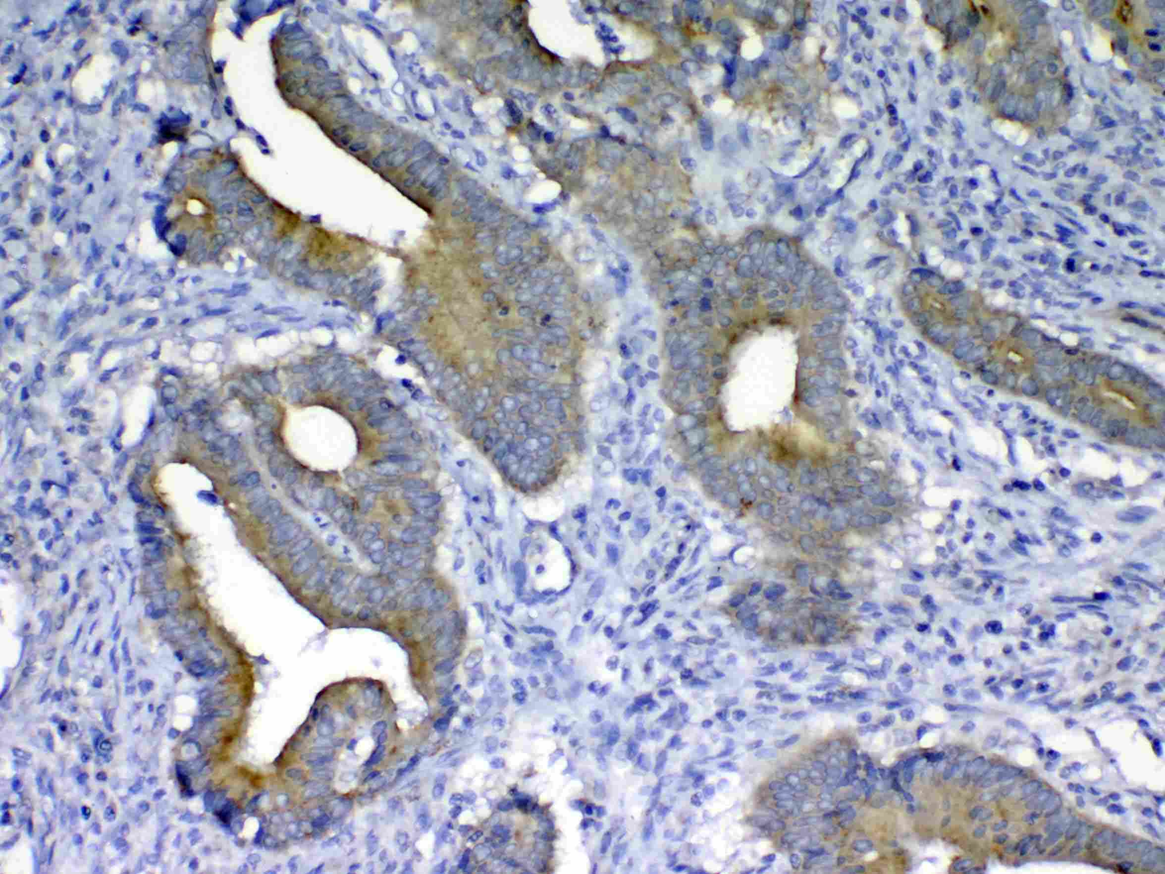
- Immunohistochemistry analysis of MMP10 on paraffin-embedded human colon cancer tissue. Antigen retrieval was performed using citrate buffer (pH6, epitope retrieval solution) for 20 mins. Sample was blocked using 10% goat serum, incubated with MMP10 polyclonal antibody (Product# PA5-79677) with a dilution of 1 µg/mL (overnight at 4°C), and followed by biotinylated goat anti-rabbit IgG (30 minutes at 37°C). Development was performed using Streptavidin-Biotin-Complex (SABC) with DAB chromogen method.
- Submitted by
- Invitrogen Antibodies (provider)
- Main image

- Experimental details
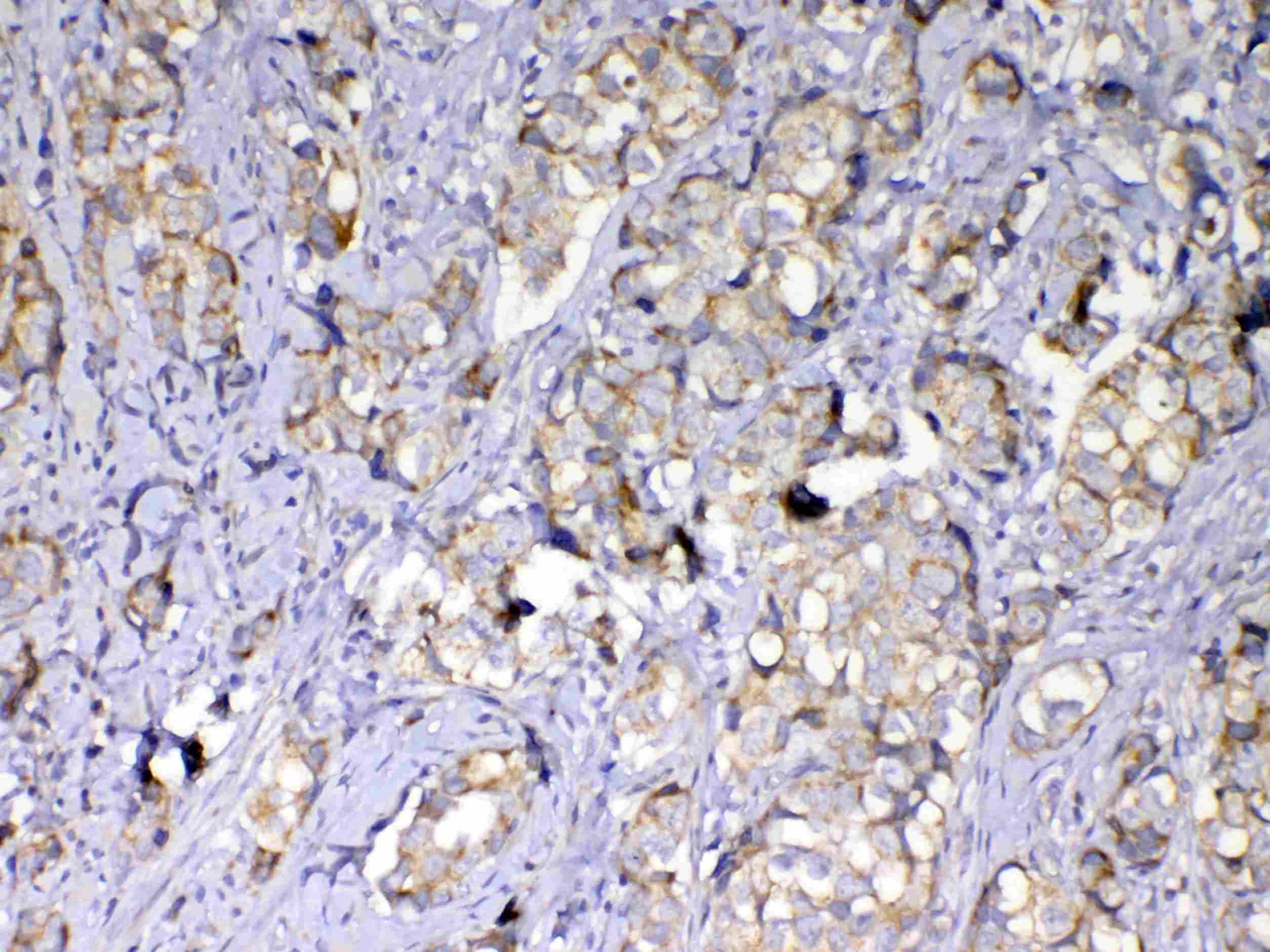
- Immunohistochemistry analysis of MMP10 on paraffin-embedded human mammary cancer tissue. Antigen retrieval was performed using citrate buffer (pH6, epitope retrieval solution) for 20 mins. Sample was blocked using 10% goat serum, incubated with MMP10 polyclonal antibody (Product# PA5-79677) with a dilution of 1 µg/mL (overnight at 4°C), and followed by biotinylated goat anti-rabbit IgG (30 minutes at 37°C). Development was performed using Streptavidin-Biotin-Complex (SABC) with DAB chromogen method.
Supportive validation
- Submitted by
- Invitrogen Antibodies (provider)
- Main image

- Experimental details
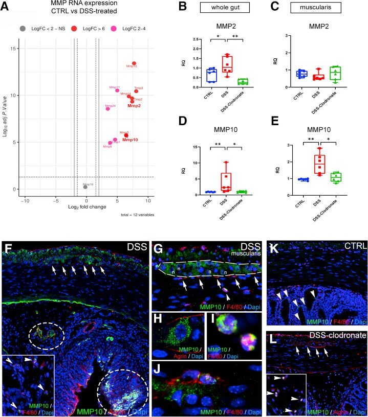
- Figure 11 Secretion of MMP10 by MMs and enteric ganglia contributes to BMB disruption in DSS-induced colitis. Volcano plot shows that MMP expression is increased in DSS-treated animals compared with controls ( A ), including MMP2 (LogFc 7.42, P < .001) and MMP10 (LogFc 6.45, P < .001). Graphs show relative RNA expression (fold change, FC) of MMP2 and MMP10 in whole gut and isolated muscularis samples. MMP2 expression is significantly increased in whole gut of DSS-treated animals ( B ) but not in the muscularis ( C ), whereas MMP10 expression is elevated in both ( D and E ). MMP10 is expressed diffusely in the longitudinal muscle layer and inside enteric ganglia after DSS treatment ( F, arrows ). In the submucosa and mucosa, MMP10 expression co-localizes with its substrate, agrin ( F, circled areas ) and F4/80+ monocytes and macrophages ( F, arrowheads ). In the muscularis, apart from enteric neurons ( G and H ), F4/80+ monocytes ( G, arrowhead ) and scattered MMs ( J ) express MMP10 but not MyMs or IGMs ( F, arrows ). In control guts, MMP10 is not expressed in the muscularis layer ( K ) but only in scattered F4/80-negative cells in the mucosa ( K, arrowheads ). In DSS+ clodronate treated animals, no MMP10 immunopositivity is detected in enteric ganglia or in the muscularis layer ( L, arrows ). In the submucosa small number of F4/80+ monocytes express MMP10 ( L, arrowheads ). Box and whisker plots (minimum-to-maximum) show median of FC values with 95 % confidence intervals. *